

Enhanced temporal variability of amygdala-frontal functional connectivity in patients with schizophrenia
- PMID: 29560309
- PMCID: PMC5857898
- DOI: 10.1016/j.nicl.2018.02.025
Enhanced temporal variability of amygdala-frontal functional connectivity in patients with schizophrenia
Abstract
Background: The "dysconnectivity hypothesis" was proposed 20 years ago. It characterized schizophrenia as a disorder with dysfunctional connectivity across a large range of distributed brain areas. Resting-state functional magnetic resonance imaging (rsfMRI) data have supported this theory. Previous studies revealed that the amygdala might be responsible for the emotion regulation-related symptoms of schizophrenia. However, conventional methods oversimplified brain activities by assuming that it remained static throughout the entire scan duration, which may explain why inconsistent results have been reported for the same brain region.
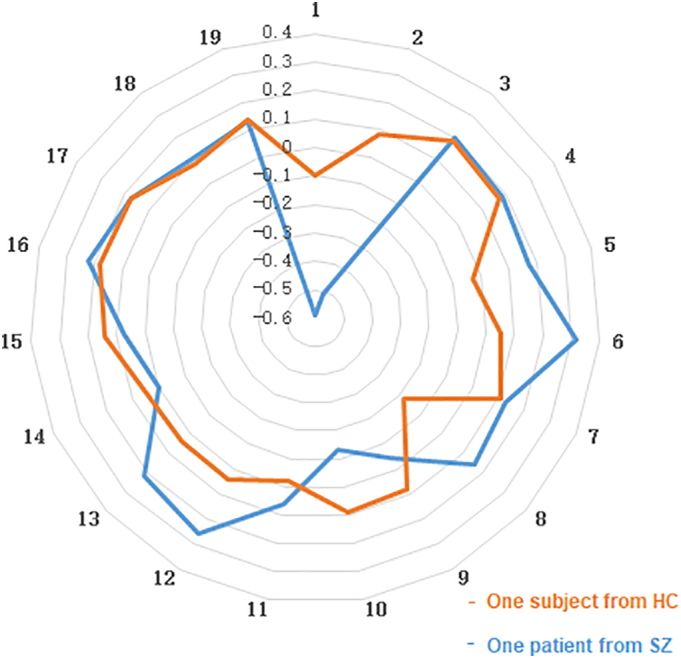
Methods: An emerging technique is sliding time window analysis, which is used to describe functional connectivity based on the temporal variability of regions of interest (e.g., amygdala) in patients with schizophrenia. Conventional analysis of the static functional connectivity between the amygdala and whole brain was also conducted.
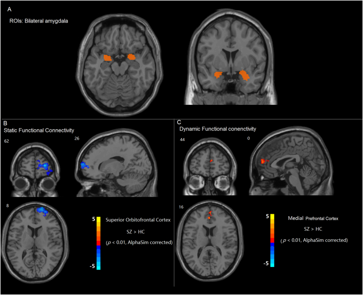
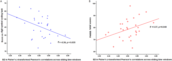
Results: Static functional connectivity between the amygdala and orbitofrontal region was impaired in patients with schizophrenia. The variability of connectivity between the amygdala and medial prefrontal cortex was enhanced (i.e., greater dynamics) in patients with schizophrenia. A negative relationship was found between the variability of connectivity and information processing efficiency. A positive correlation was found between the variability of connectivity and symptom severity.
Conclusion: The findings suggest that schizophrenia was related to abnormal patterns of fluctuating communication among brain areas that are involved in emotion regulations. Unveiling the temporal properties of functional connectivity could disentangle the inconsistent results of previous functional connectivity studies.
Keywords: Amygdala; Dynamic functional connectivity; Schizophrenia; Sliding-window correlation analysis; Temporal variability.
Figures
Similar articles
-
Abnormal effective fronto-limbic connectivity during emotion processing in schizophrenia.Prog Neuropsychopharmacol Biol Psychiatry. 2017 Jan 4;72:1-8. doi: 10.1016/j.pnpbp.2016.08.004. Epub 2016 Aug 12. Prog Neuropsychopharmacol Biol Psychiatry. 2017. PMID: 27528110
-
Altered amygdala-prefrontal connectivity during emotion perception in schizophrenia.Schizophr Res. 2016 Aug;175(1-3):35-41. doi: 10.1016/j.schres.2016.04.003. Epub 2016 Apr 12. Schizophr Res. 2016. PMID: 27083779
-
Incapacity to control emotion in major depression may arise from disrupted white matter integrity and OFC-amygdala inhibition.CNS Neurosci Ther. 2018 Nov;24(11):1053-1062. doi: 10.1111/cns.12800. Epub 2018 Jan 24. CNS Neurosci Ther. 2018. PMID: 29368421 Free PMC article.
-
Frontal lobe alterations in schizophrenia: a review.Trends Psychiatry Psychother. 2016 Oct-Dec;38(4):198-206. doi: 10.1590/2237-6089-2015-0088. Trends Psychiatry Psychother. 2016. PMID: 28076640 Review.
-
Resting-state functional MRI in treatment-resistant schizophrenia.Front Neuroimaging. 2023 Apr 6;2:1127508. doi: 10.3389/fnimg.2023.1127508. eCollection 2023. Front Neuroimaging. 2023. PMID: 37554635 Free PMC article. Review.
Cited by
-
Test-retest reliability of time-varying patterns of brain activity across single band and multiband resting-state functional magnetic resonance imaging in healthy older adults.Front Hum Neurosci. 2022 Nov 10;16:980280. doi: 10.3389/fnhum.2022.980280. eCollection 2022. Front Hum Neurosci. 2022. PMID: 36438643 Free PMC article.
-
Dynamic Functional Connectivity Reveals Abnormal Variability in the Amygdala Subregions of Children With Attention-Deficit/Hyperactivity Disorder.Front Neurosci. 2021 Sep 29;15:648143. doi: 10.3389/fnins.2021.648143. eCollection 2021. Front Neurosci. 2021. PMID: 34658751 Free PMC article.
-
Neuroimage Analysis Methods and Artificial Intelligence Techniques for Reliable Biomarkers and Accurate Diagnosis of Schizophrenia: Achievements Made by Chinese Scholars Around the Past Decade.Schizophr Bull. 2025 Mar 14;51(2):325-342. doi: 10.1093/schbul/sbae110. Schizophr Bull. 2025. PMID: 38982882 Free PMC article. Review.
-
Functional connectivity between the amygdala and prefrontal cortex underlies processing of emotion ambiguity.Transl Psychiatry. 2023 Oct 28;13(1):334. doi: 10.1038/s41398-023-02625-w. Transl Psychiatry. 2023. PMID: 37898626 Free PMC article.
-
Tools of the trade: estimating time-varying connectivity patterns from fMRI data.Soc Cogn Affect Neurosci. 2021 Aug 5;16(8):849-874. doi: 10.1093/scan/nsaa114. Soc Cogn Affect Neurosci. 2021. PMID: 32785604 Free PMC article. Review.
References
Publication types
MeSH terms
LinkOut - more resources
Full Text Sources
Other Literature Sources
Medical
