. 2018 Jan 22;5(3):297-314.
doi: 10.1002/acn3.527. eCollection 2018 Mar.

Targeting potassium channels to treat cerebellar ataxia

Affiliations

Targeting potassium channels to treat cerebellar ataxia

David D Bushart et al. Ann Clin Transl Neurol. .

Abstract

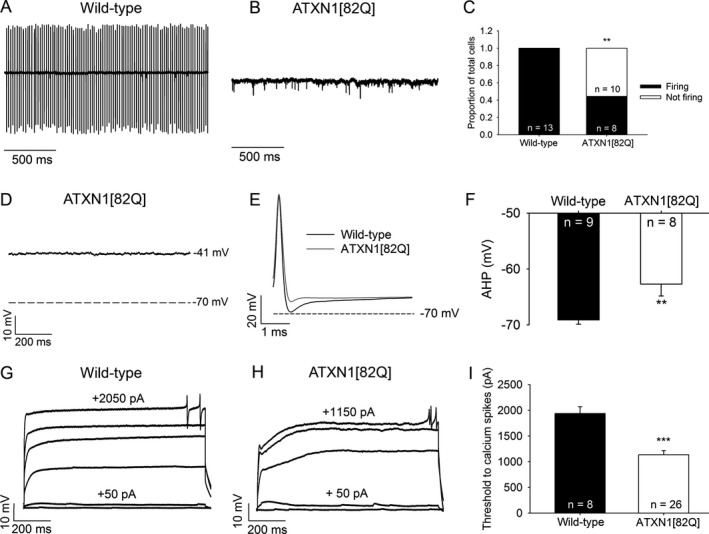
Objective: Purkinje neuron dysfunction is associated with cerebellar ataxia. In a mouse model of spinocerebellar ataxia type 1 (SCA1), reduced potassium channel function contributes to altered membrane excitability resulting in impaired Purkinje neuron spiking. We sought to determine the relationship between altered membrane excitability and motor dysfunction in SCA1 mice.

Methods: Patch-clamp recordings in acute cerebellar slices and motor phenotype testing were used to identify pharmacologic agents which improve Purkinje neuron physiology and motor performance in SCA1 mice. Additionally, we retrospectively reviewed records of patients with SCA1 and other autosomal-dominant SCAs with prominent Purkinje neuron involvement to determine whether currently approved potassium channel activators were tolerated.

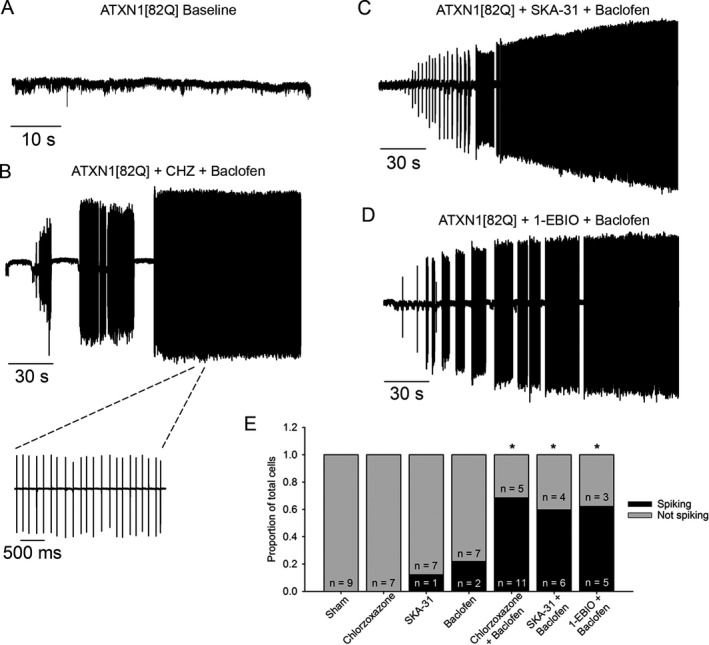
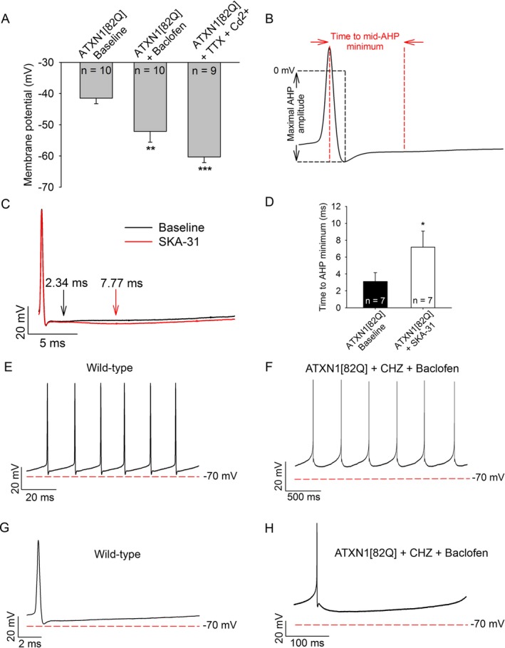
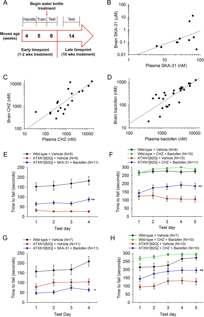
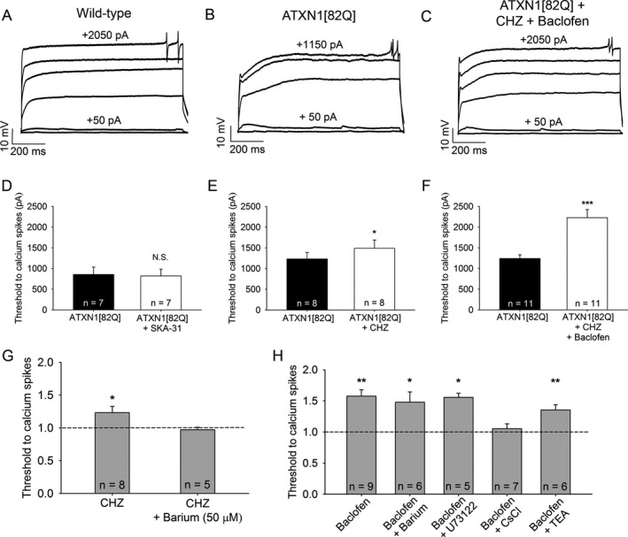
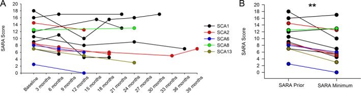
Results: Activating calcium-activated and subthreshold-activated potassium channels improved Purkinje neuron spiking impairment in SCA1 mice (P < 0.05). Additionally, dendritic hyperexcitability was improved by activating subthreshold-activated potassium channels but not calcium-activated potassium channels (P < 0.01). Improving spiking and dendritic hyperexcitability through a combination of chlorzoxazone and baclofen produced sustained improvements in motor dysfunction in SCA1 mice (P < 0.01). Retrospective review of SCA patient records suggests that co-treatment with chlorzoxazone and baclofen is tolerated.

Interpretation: Targeting both altered spiking and dendritic membrane excitability is associated with sustained improvements in motor performance in SCA1 mice, while targeting altered spiking alone produces only short-term improvements in motor dysfunction. Potassium channel activators currently in clinical use are well tolerated and may provide benefit in SCA patients. Future clinical trials with potassium channel activators are warranted in cerebellar ataxia.


Figures

Figure 1
Figure 1
ATXN1[82Q] Purkinje neurons display both an absence of repetitive spiking and dendritic hyperexcitability. (A) Representative spiking of a wild‐type Purkinje neuron in the cell‐attached recording configuration. (B) Representative trace of a nonspiking ATXN1[82Q] Purkinje neuron in the cell‐attached recording configuration. (C) Summary of spiking and nonspiking Purkinje neurons from wild‐type and ATXN1[82Q] mice. (D) Representative trace of a nonfiring ATXN1[82Q] Purkinje neuron in the whole‐cell recording configuration. These neurons display a depolarized resting membrane potential. (E) After‐hyperpolarization (AHP) amplitude in wild‐type and ATXN1[82Q] Purkinje neurons. (F) Summary of AHP amplitudes in wild‐type and ATXN1[82Q] Purkinje neurons. (G) Representative trace of a wild‐type Purkinje neuron held at −80 mV in the presence of tetrodotoxin. Upon injection of positive current in +50 pA increments, dendritic calcium spikes are noted. (H) Representative trace of dendritic calcium spike analysis from an ATXN1[82Q] Purkinje neuron. (I) Summary of the threshold of injected current required to elicit dendritic calcium spikes in wild‐type and ATXN1[82Q] Purkinje neurons in the presence of tetrodotoxin. **P < 0.01, ***P < 0.001, Fisher's exact test (C) or two‐sample Student's t‐test (I).
Figure 2
Figure 2
Potassium channel‐activating compounds restore spiking in nonfiring ATXN1[82Q] Purkinje neurons. (A) In a cell‐attached recording configuration, the majority of ATXN1[82Q] Purkinje neurons are nonfiring at 5 weeks of age. (B) Co‐application of chlorzoxazone (CHZ, 50 μmol/L) and baclofen (10 μmol/L) restores repetitive spiking to nonfiring ATXN1[82Q] Purkinje neurons (P = 0.001). Inset of restored spiking with chlorzoxazone and baclofen is shown on an expanded time scale. (C) SKA‐31 (10 μmol/L) and baclofen (10 μmol/L) co‐application also restores spiking to nonfiring ATXN1[82Q] Purkinje neurons (P = 0.01), as does (D) 1‐EBIO (100 μmol/L) and baclofen (10 μmol/L) (P = 0.009). (E) Summary of data from figures B–D. *adjusted P < 0.01 when compared to sham, Fisher's exact test with Bonferroni postcorrection.
Figure 3
Figure 3
KC a activators and baclofen enhance the AHP and repolarize the membrane potential of ATXN1[82Q] Purkinje neurons. (A) Baclofen (10 μmol/L) hyperpolarizes the membrane potential of depolarized ATXN1[82Q] Purkinje neurons to from −41 mV to −52 mV. Tetrodotoxin (1 μmol/L) and cadmium (100 μmol/L) repolarizes the membrane potential to −60 mV. (B) Protocol for analysis of the time to minimal mid‐AHP and maximal AHP amplitude. (C) Representative trace of the AHP of an ATXN1[82Q] Purkinje neuron before (black trace) and after (red trace) SKA‐31 perfusion (10 μmol/L). The time to slow AHP minimum is denoted by arrows. (D) Summary of data from panel C. SKA‐31 extends the duration of the AHP in ATXN1[82Q] Purkinje neurons (P = 0.042). (E) Representative trace which displays the interspike interval during spontaneous firing of a baseline wild‐type Purkinje neuron and (F) ATXN1[8Q] Purkinje neuron in the presence of chlorzoxazone (50 μmol/L) and baclofen (10 μmol/L). (G) Single interspike intervals of baseline wild‐type and (H) ATXN1[82Q] Purkinje neurons in the presence of chlorzoxazone and baclofen. *P < 0.05, **P < 0.01, ***P < 0.001, paired Student's t‐test. CHZ, chlorzoxazone.
Figure 4
Figure 4
Chlorzoxazone and baclofen, but not SKA‐31 and baclofen, sustains improvement in motor dysfunction in ATXN1[82Q] mice. (A) Drug administration and behavioral testing paradigm. (B) Correlated brain and plasma levels of SKA‐31 are seen after administration through drinking water (R 2 = 0.1337). (C) Correlated brain and plasma levels of chlorzoxazone are seen after administration through drinking water (R 2 = 0.8904). (D) Correlated brain and plasma levels of baclofen are present after administration through drinking water (R 2 = 0.8591). (E) After 1 week of treatment, SKA‐31 + baclofen improves motor performance in ATXN1[82Q] mice (F(2, 113) = 15.76, P < 0.0001) (Wild‐type + Vehicle vs. ATXN1[82Q] + Vehicle P < 0.0001; Wild‐type + Vehicle vs. ATXN1[82Q] + SKA‐31 + Baclofen P < 0.0001; ATXN1[82Q] + Vehicle vs. ATXN1[82Q] + SKA‐31 + Baclofen P = 0.004). (F) After 1 week of treatment, chlorzoxazone + baclofen improves motor performance in ATXN1[82Q] mice (F(3, 156) = 42.23, P < 0.0001) (Wild‐type + Vehicle vs. Wild‐type + Chlorzoxazone + Baclofen P = 0.9726; Wild‐type + Vehicle vs. ATXN1[82Q] + Vehicle P < 0.0001; Wild‐type + Vehicle vs. ATXN1[82Q] + Chlorzoxazone + Baclofen P < 0.0001; Wild‐type + Chlorzoxazone + Baclofen vs. ATXN1[82Q] + Vehicle P < 0.0001; Wild‐type + Chlorzoxazone + Baclofen vs. ATXN1[82Q] + Chlorzoxazone + Baclofen P < 0.0001; ATXN1[82Q] + Vehicle vs. ATXN1[82Q] + Chlorzoxazone + Baclofen P = 0.0036). (G) After 10 weeks of treatment, mice treated with SKA‐31+ baclofen show worsened motor performance compared with vehicle‐treated controls (F(2, 109) = 36.73, P < 0.0001) (Wild‐type vs. ATXN1[82Q] + Vehicle P = 0.0005; Wild‐type vs. ATXN1[82Q] + SKA‐31 + Baclofen P < 0.0001; ATXN1[82Q] + Vehicle vs. ATXN1[82Q] + SKA‐31 + Baclofen P = 0.0408). (H) After 10 weeks of treatment, ATXN1[82Q] mice treated with chlorzoxazone + baclofen display sustained improvement in motor performance compared with vehicle‐treated controls (F(3, 144) = 29.43, P < 0.0001) (Wild‐type + Vehicle vs. Wild‐type + Chlorzoxazone + Baclofen P = 0.0292; Wild‐type + Vehicle vs. ATXN1[82Q] + Vehicle P < 0.0001; Wild‐type + Vehicle vs. ATXN1[82Q] + Chlorzoxazone + Baclofen P = 0.0097; Wild‐type + Chlorzoxazone + Baclofen vs. ATXN1[82Q] + Vehicle P < 0.0001; Wild‐type + Chlorzoxazone + Baclofen vs. ATXN1[82Q] + Chlorzoxazone + Baclofen P < 0.0001; ATXN1[82Q] + Vehicle vs. ATXN1[82Q] + Chlorzoxazone + Baclofen P = 0.0029). *P < 0.05, ** P < 0.01, two‐way ANOVA with Holm–Sidak posttest. CHZ, chlorzoxazone.
Figure 5
Figure 5
Chlorzoxazone and baclofen reduce dendritic hyperexcitability in ATXN1[82Q] mice by activating subthreshold‐activated potassium channels. (A) Representative trace of dendritic calcium spikes from a wild‐type Purkinje neuron, (B) ATXN1[82Q] Purkinje neuron at baseline, and (C) the same ATXN1[82Q] Purkinje neuron treated with chlorzoxazone (50 μmol/L) and baclofen (2 μmol/L). (D) SKA‐31 (10 μmol/L) does not reduce dendritic hyperexcitability in ATXN1[82Q] Purkinje neurons (P = 0.376). (E) Chlorzoxazone (50 μmol/L) reduces dendritic hyperexcitability in ATXN1[82Q] Purkinje neurons (P = 0.025). (F) Chlorzoxazone (50 μmol/L) and baclofen (2 μmol/L) coadministration further reduces dendritic excitability in ATXN1[82Q] Purkinje neurons (P < 0.001). (G) Barium (50 μmol/L) occludes the effect of chlorzoxazone on dendritic excitability (P = 0.778). (H) Barium (500 μmol/L, P = 0.012), U73122 (10 μmol/L in recording pipette, P = 0.014), and TEA (1 mmol/L, P = 0.009) do not occlude the effect of baclofen on dendritic excitability, but cesium chloride (140 mmol/L in the recording pipette) does occlude the effect of baclofen on dendritic excitability (P = 0.356) in ATXN1[82Q] Purkinje neurons. *P < 0.05, **P < 0.01, ***P < 0.001, paired Student's t‐test. CHZ, chlorzoxazone.
Figure 6
Figure 6
Chlorzoxazone and baclofen coadministration is tolerated in spinocerebellar ataxia patients and improves symptoms. (A) SARA scores were obtained for each patient prior to beginning treatment with chlorzoxazone and baclofen, and subsequent SARA scores were obtained at follow‐up visits. SARA scores are only displayed for patients who could tolerate treatment and had at least one follow‐up visit. (B) SARA scores are displayed prior to treatment and at the time point which showed a minimum SARA score after beginning treatment (P = 0.004). **P < 0.01, paired Student's t‐test.

Similar articles

Cited by

References

    1. Dell'Orco JM, Pulst SM, Shakkottai VG. Potassium channel dysfunction underlies Purkinje neuron spiking abnormalities in spinocerebellar ataxia type 2. Hum Mol Genet 2017;15:3935–3945. - PMC - PubMed
    1. Dell'Orco JM, Wasserman AH, Chopra R, et al. Neuronal Atrophy Early in Degenerative Ataxia Is a Compensatory Mechanism to Regulate Membrane Excitability. J Neurosci 2015;12(35):11292–11307. - PMC - PubMed
    1. Shakkottai VG, do Carmo Costa M, Dell'Orco JM, et al. Early changes in cerebellar physiology accompany motor dysfunction in the polyglutamine disease spinocerebellar ataxia type 3. J Neurosci 2011;31:13002–13014. - PMC - PubMed
    1. Hansen ST, Meera P, Otis TS, Pulst SM. Changes in Purkinje cell firing and gene expression precede behavioral pathology in a mouse model of SCA2. Hum Mol Genet 2013;22:271–283. - PMC - PubMed
    1. Ferrer I, Genis D, Davalos A, et al. The Purkinje cell in olivopontocerebellar atrophy. A Golgi and immunocytochemical study. Neuropathol Appl Neurobiol 1994;20:38–46. - PubMed