

doi: 10.1212/NXG.0000000000000226.
eCollection 2018 Apr.
Whole-exome sequencing identifies mutations in MYMK in a mild form of Carey-Fineman-Ziter syndrome
Affiliations
- PMID: 29560417
- PMCID: PMC5858950
- DOI: 10.1212/NXG.0000000000000226
Item in Clipboard
Whole-exome sequencing identifies mutations in MYMK in a mild form of Carey-Fineman-Ziter syndrome
Neurol Genet.
.
No abstract available
Figures
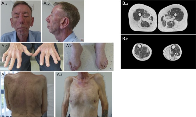
(A.a and A.b) Front and profile facial photographs demonstrating lagophthalmos (A.a, patient attempting lid closure), muscle hypoplasia, retrognathia, and broad nasal tip. (A.c and A.d) Wasting of intrinsic hand muscles and contracture deformities of the right little finger and the toes. (A.e, A.f) Scoliosis (A.e) and generalized muscle atrophy with pectoralis muscle hypoplasia (A.f). (B) T2-weighted MRIs of the thighs (B.a) showing severe fatty replacement of hamstrings, thigh adductors, and sartorius muscles, with relative sparing of the gracilis and quadriceps muscles bilaterally, and of the calves (B.b) showing asymmetric involvement with more marked fatty replacement in muscle of the right leg. Gastrocnemius and soleus muscles are severely affected, and the tibialis anterior on the right is relatively spared.
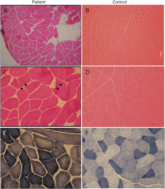
Patient images and control images. H&E stain demonstrates fiber-size variation (A and B; H&E ×100) and occasional internal nuclei (C and D; H&E ×200, arrows). Mild moth-eaten changes seen on nicotinamide adenine dinucleotide (NADH) stain, indicating uneven mitochondrial enzyme activity within the sarcoplasm (E and F; NADH ×200).
References
-
- Zhang W, Roy S. Myomaker is required for the fusion of fast-twitch myocytes in the zebrafish embryo. Dev Biol 2017;423:24–33. - PubMed
Grants and funding
LinkOut - more resources
Full Text Sources
Other Literature Sources