

Distribution of Interleukin-22-secreting Immune Cells in Conjunctival Associated Lymphoid Tissue
- PMID: 29560621
- PMCID: PMC5906400
- DOI: 10.3341/kjo.2017.0068
Distribution of Interleukin-22-secreting Immune Cells in Conjunctival Associated Lymphoid Tissue
Abstract
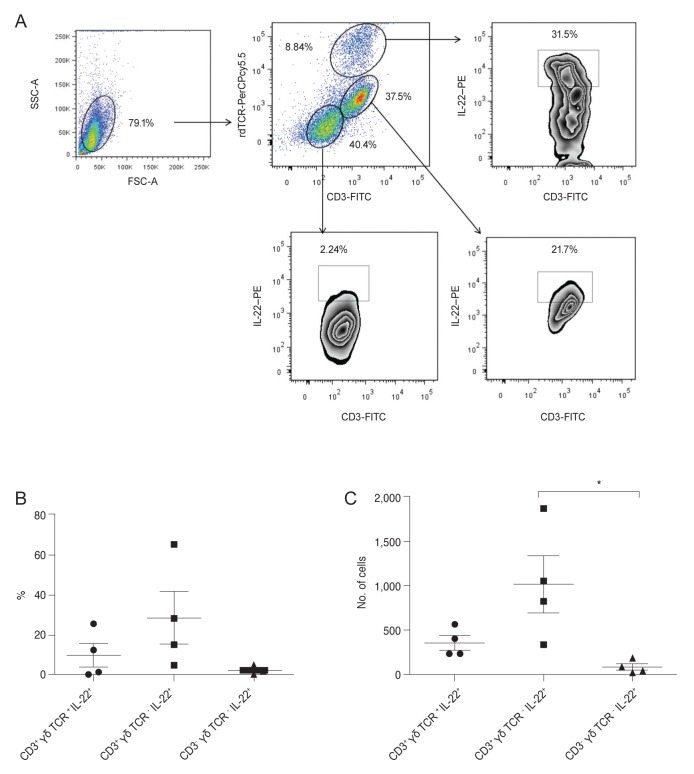
Purpose: Interleukin (IL)-22 is a cytokine involved in epithelial cell regeneration. Currently, no research studies have analyzed the distribution of the three distinct IL-22-secreting cell populations in human or mouse conjunctiva. This study investigated the distribution of the three main populations of IL-22-secreting immune cells, αβ Th cells, γδ T cells, or innate cells (innate lymphoid cells [ILCs] or natural killer cells), in conjunctival associated lymphoid tissues (CALTs) in human and mouse models.
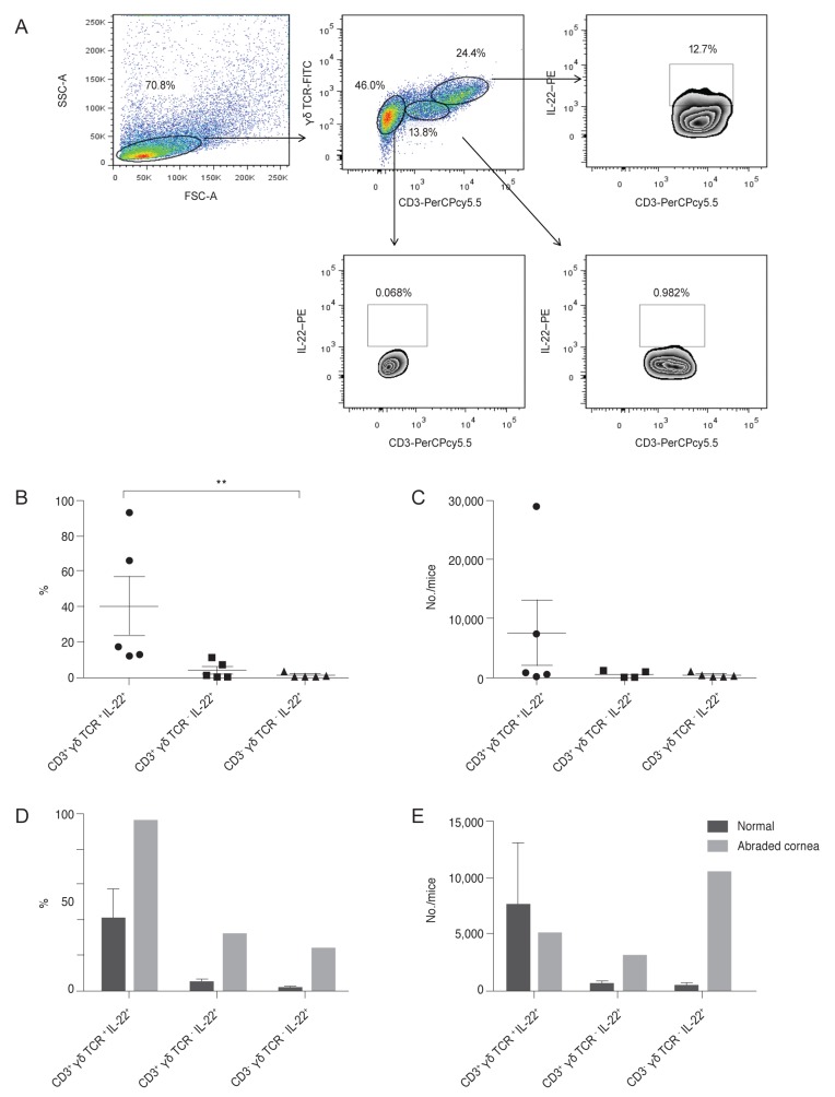
Methods: We collected discarded cadaveric bulbar conjunctival tissue specimens after preservation of the corneo-limbal tissue for keratoplasty from four enucleated eyes of the domestic donor. The bulbar conjunctiva tissue, including the cornea from normal (n = 27) or abraded (n = 4) B6 mice, were excised and pooled in RPMI 1640 media. After the lymphoid cells were gated in forward and side scattering, the αβ Th cells, γδ T cells, or innate lymphoid cells were positively or negatively gated using anti-CD3, anti-γδ TCR, and anti-IL-22 antibodies, with a FACSCanto flow cytometer.
Results: In normal human conjunctiva, the percentage and number of cells were highest in αβ Th cells, followed by γδ T cells and CD3- γδ TCR- IL-22+ innate cells (presumed ILCs, pILCs) (Kruskal-Wallis test, p = 0.012). In normal mice keratoconjunctiva, the percentage and total number were highest in γδ T cells, followed by αβ Th cells and pILCs (Kruskal-Wallis test, p = 0.0004); in corneal abraded mice, the population of αβ Th cells and pILCs tended to increase.
Conclusions: This study suggests that three distinctive populations of IL-22-secreting immune cells are present in CALTs of both humans and mice, and the proportions of IL-22+αβ Th cells, γδ T cells, and pILCs in CALTs in humans might be differently distributed from those in normal mice.
Keywords: Conjunctiva; Conjunctival associated lymphoid tissues; Innate lymphoid cell; Interleukin-22; Th22 cell.
© 2018 The Korean Ophthalmological Society.
Conflict of interest statement
No potential conflict of interest relevant to this article was reported.
Figures
Similar articles
-
Whodunit? The Contribution of Interleukin (IL)-17/IL-22-Producing γδ T Cells, αβ T Cells, and Innate Lymphoid Cells to the Pathogenesis of Spondyloarthritis.Front Immunol. 2018 Apr 25;9:885. doi: 10.3389/fimmu.2018.00885. eCollection 2018. Front Immunol. 2018. PMID: 29922283 Free PMC article. Review.
-
Mice depleted of alphabeta but not gammadelta T cells are resistant to mortality caused by cecal ligation and puncture.Shock. 2007 May;27(5):507-19. doi: 10.1097/SHK.0b013e31802b5d9f. Shock. 2007. PMID: 17438456
-
Epithelial component and intraepithelial lymphocytes of conjunctiva-associated lymphoid tissue in healthy children.Histol Histopathol. 2021 Dec;36(12):1273-1283. doi: 10.14670/HH-18-385. Epub 2021 Oct 26. Histol Histopathol. 2021. PMID: 34698365
-
{gamma}{delta} T cell homeostasis is established in competition with {alpha}{beta} T cells and NK cells.Proc Natl Acad Sci U S A. 2005 Oct 11;102(41):14741-6. doi: 10.1073/pnas.0507520102. Epub 2005 Oct 3. Proc Natl Acad Sci U S A. 2005. PMID: 16203967 Free PMC article.
-
Human γδ T-Cells: From Surface Receptors to the Therapy of High-Risk Leukemias.Front Immunol. 2018 May 7;9:984. doi: 10.3389/fimmu.2018.00984. eCollection 2018. Front Immunol. 2018. PMID: 29867961 Free PMC article. Review.
Cited by
-
Plasmacytoid dendritic cells in the eye.Prog Retin Eye Res. 2021 Jan;80:100877. doi: 10.1016/j.preteyeres.2020.100877. Epub 2020 Jul 24. Prog Retin Eye Res. 2021. PMID: 32717378 Free PMC article. Review.
-
Double-edged sword: γδ T cells in mucosal homeostasis and disease.Exp Mol Med. 2023 Sep;55(9):1895-1904. doi: 10.1038/s12276-023-00985-3. Epub 2023 Sep 11. Exp Mol Med. 2023. PMID: 37696894 Free PMC article. Review.
-
Conjunctival Intraepithelial Lymphocytes, Lacrimal Cytokines and Ocular Commensal Microbiota: Analysis of the Three Main Players in Allergic Conjunctivitis.Front Immunol. 2022 Jul 19;13:911022. doi: 10.3389/fimmu.2022.911022. eCollection 2022. Front Immunol. 2022. PMID: 35935953 Free PMC article.
-
Differentiation and regulation of CD4+ T cell subsets in Parkinson's disease.Cell Mol Life Sci. 2024 Aug 17;81(1):352. doi: 10.1007/s00018-024-05402-0. Cell Mol Life Sci. 2024. PMID: 39153043 Free PMC article. Review.
-
The ocular surface immune system through the eyes of aging.Ocul Surf. 2021 Apr;20:139-162. doi: 10.1016/j.jtos.2021.02.007. Epub 2021 Feb 20. Ocul Surf. 2021. PMID: 33621658 Free PMC article. Review.
References
Publication types
MeSH terms
Substances
LinkOut - more resources
Full Text Sources
Other Literature Sources
