. 2018 Mar;23(3):1-4.
doi: 10.1117/1.JBO.23.3.030504.

Radiotherapy-induced Cherenkov luminescence imaging in a human body phantom

Affiliations

Radiotherapy-induced Cherenkov luminescence imaging in a human body phantom

Syed Rakin Ahmed et al. J Biomed Opt. 2018 Mar.

Abstract

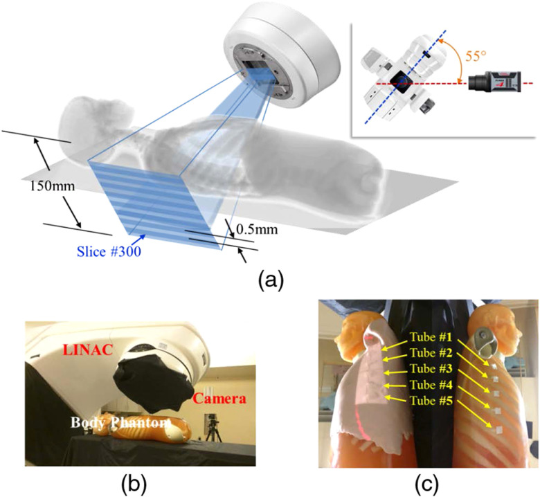
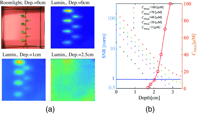
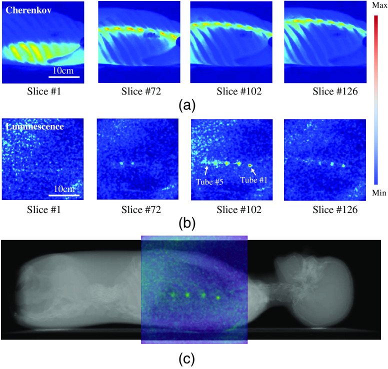
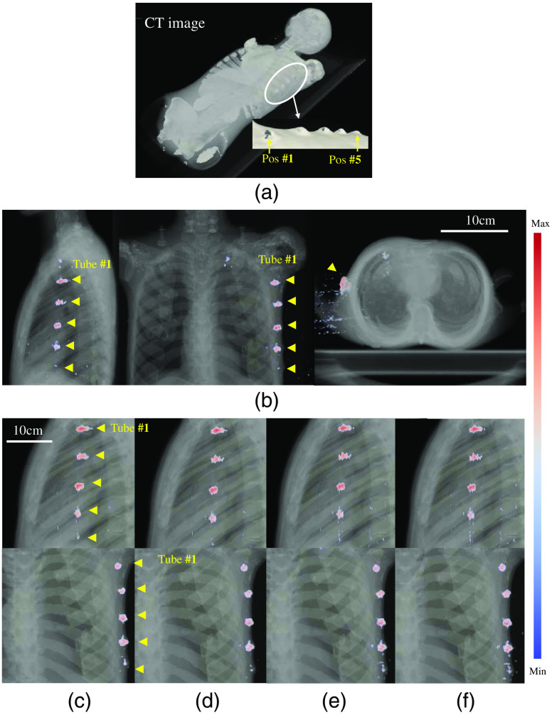
Radiation therapy produces Cherenkov optical emission in tissue, and this light can be utilized to activate molecular probes. The feasibility of sensing luminescence from a tissue molecular oxygen sensor from within a human body phantom was examined using the geometry of the axillary lymph node region. Detection of regions down to 30-mm deep was feasible with submillimeter spatial resolution with the total quantity of the phosphorescent sensor PtG4 near 1 nanomole. Radiation sheet scanning in an epi-illumination geometry provided optimal coverage, and maximum intensity projection images provided illustration of the concept. This work provides the preliminary information needed to attempt this type of imaging in vivo.

Keywords: Cerenkov; linac; phosphorescence; radiation; therapy.


Figures

Fig. 1
Fig. 1
Measurement geometry for the body phantom is shown with (a) a schematic of the geometry, (b) a photograph of the setup, and (c) the body phantom with skin on (left) and without skin (right) as shown with five tubes fixed onto the lateral rib area.
Fig. 2
Fig. 2
Experimental results to investigate image quality versus depth for different concentrations of PtG4 (CPtG4) as shown in the legend, showing (a) the room light image and luminescence images of the phantom used at three different depths in the phantom, and (b) the corresponding SNR values extracted from these images are shown as a function of depth by colored dots. In the same graph, the red line shows the CPtG4 at which SNR=1 (values on left y-axis).
Fig. 3
Fig. 3
Raw measurement sequences are shown for (a) Cherenkov emission and (b) luminescence photons, for the sheet at four different slice locations. In (c) a composition image is shown of the phantom CT with color overlay of the CELSI MIP image. A temporal sweep of the signals in (a) and (b) can be seen in the associated video files as the sheet moved across the phantom body.
Fig. 4
Fig. 4
3-D images of (a) CT and (b) overlaid with CELSI for three orthogonal views: coronal, sagittal, and transverse. Compositions of CT and CELSI images for varying numbers of light sheets, which correspond to less dose delivery. In these images, the number of sheets were: (c) 25, (d) 50, (e) 100, and (f) 250, respectively.

References

    1. Razansky D., et al. , “Deep tissue optical and optoacoustic molecular imaging technologies for pre-clinical research and drug discovery,” Curr. Pharm. Biotechnol. 13(4), 504–522 (2012).10.2174/138920112799436258 - DOI - PubMed
    1. Zhang R., et al. , “Cherenkov-excited luminescence scanned imaging,” Opt. Lett. 40(5), 827–830 (2015).OPLEDP10.1364/OL.40.000827 - DOI - PubMed
    1. Jarvis L. A., et al. , “Cherenkov video imaging allows for the first visualization of radiation therapy in real time,” Int. J. Radiat. Oncol. Biol. Phys. 89(3), 615–622 (2014).IOBPD310.1016/j.ijrobp.2014.01.046 - DOI - PubMed
    1. Robertson R., et al. , “Optical imaging of Cerenkov light generation from positron-emitting radiotracers,” Phys. Med. Biol. 54(16), N355–N365 (2009).PHMBA710.1088/0031-9155/54/16/N01 - DOI - PMC - PubMed
    1. Axelsson J., et al. , “Quantitative Cherenkov emission spectroscopy for tissue oxygenation assessment,” Opt. Express 20(5), 5133–5142 (2012).OPEXFF10.1364/OE.20.005133 - DOI - PMC - PubMed

Publication types