

Cerebrovascular blood oxygenation level dependent pulsatility at baseline and following acute exercise among healthy adolescents
- PMID: 29561225
- PMCID: PMC6727139
- DOI: 10.1177/0271678X18766771
Cerebrovascular blood oxygenation level dependent pulsatility at baseline and following acute exercise among healthy adolescents
Abstract
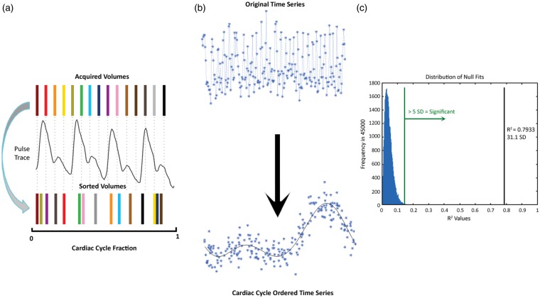
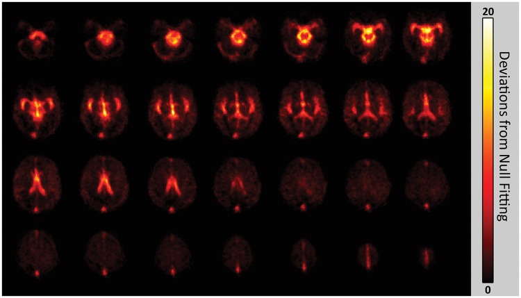
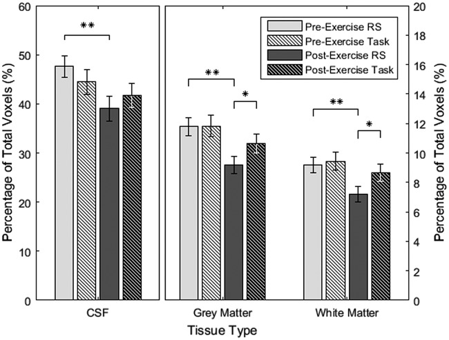
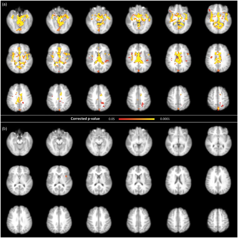
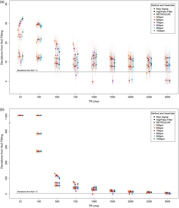
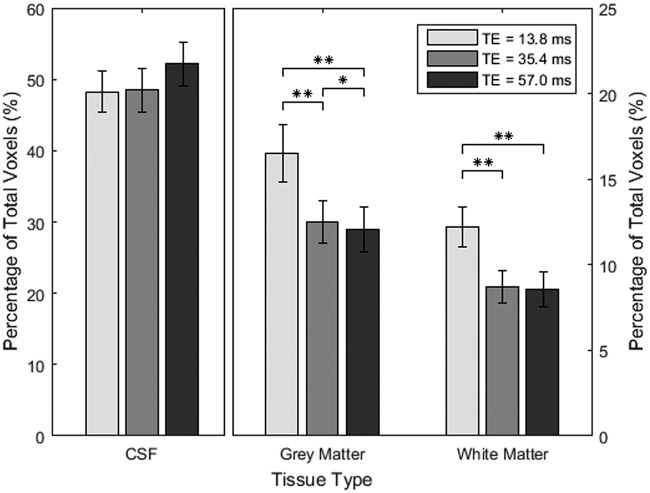
Arterial stiffness is linked to cerebral small vessel damage and neurodegeneration, but barriers to accessing deep cerebrovascular anatomy limit our ability to assess the brain. This study describes an adaptation of a cardiac-related scrubbing method as a means of generating blood oxygenation level-dependent pulsatility maps based on the cardiac cycle. We examine BOLD pulsatility at rest, based on the non-parametric deviation from null metric, as well as changes following acute physiological stress from 20 min of moderate-intensity cycling in 45 healthy adolescents. We evaluate the influence of repetition time (TR) and echo time (TE) using simulated and multi-echo empirical data, respectively. There were tissue-specific and voxel-wise BOLD pulsatility decreases 20 min following exercise cessation. BOLD pulsatility detection was comparable over a range of TR and TE values when scan volumes were kept constant; however, short TRs (≤500 ms) and TEs (∼14 ms) acquisitions would yield the most efficient detection. Results suggest cardiac-related BOLD pulsatility may represent a robust and easily adopted method of mapping cerebrovascular pulsatility with voxel-wise resolution.
Keywords: Functional magnetic resonance imaging; aerobic exercise; blood oxygenation level dependent signal; cardiac cycle; cerebrovascular pulsatility; physiological fluctuations.
Figures
References
-
- Cipolla MJ. Control of cerebral blood flow. In: Granger and Granger (eds) The cerebral circulation. San Rafael, CA: Morgan & Claypool Life Scienceshttp, 2009, pp.27–32.
-
- O’Rourke MF, Hashimoto J. Mechanical factors in arterial aging. J Am Coll Cardiol 2007; 50: 1–13. - PubMed
-
- Webb AJS, Simoni M, Mazzucco S, et al. Increased cerebral arterial pulsatility in patients with leukoaraiosis: arterial stiffness enhances transmission of aortic pulsatility. Stroke 2012; 43: 2631–2636. - PubMed
Publication types
MeSH terms
Substances
LinkOut - more resources
Full Text Sources
Other Literature Sources
Medical
