

Correlation between panoramic radiography and cone-beam computed tomography in assessing maxillary impacted canines
- PMID: 29561656
- PMCID: PMC8191926
- DOI: 10.2319/103117-739.1
Correlation between panoramic radiography and cone-beam computed tomography in assessing maxillary impacted canines
Abstract
Objective: To determine the usefulness of panoramic radiographs in determining the labio-palatal position of maxillary impacted canines (MICs) and root resorption of permanent incisors on cone-beam computed tomography (CBCT) in correlation with the mesiodistal position of MICs on panoramic radiographs.
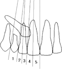
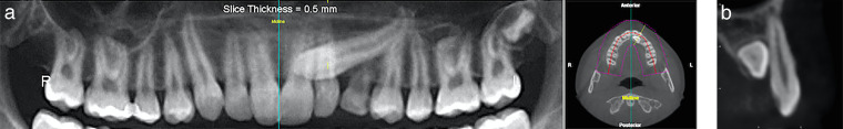
Materials and methods: This retrospective radiographic study reviewed 64 patients with 86 MICs. Subjects were divided into two groups: group I (<15 years old) and group II (>15 years old). The mesiodistal position of the MICs' cusp tips was classified into five sectors on panoramic radiographs. The labio-palatal position of the MICs and root resorption of permanent incisors were evaluated on CBCT. The statistical correlation between panoramic radiograph and CBCT results was examined using the chi-square test and the Fisher exact test.
Results: Most of the positions of MICs were palatal (67%), followed by labial (28%) and mid-alveolus (5%; P < .05). Labial positioned MICs on CBCT were more frequent in panoramic sector 1, mid-alveolus MICs were more common in sector 2, and palatally positioned MICs were more frequent in sectors 3, 4, and 5. The association between sectors of the MICs on panoramic images and the labio-palatal position of the MICs on CBCT was statistically significant ( P < .001). Root resorption of adjacent incisors showed a significant difference according to sector location ( P < .01) and was mainly found in sectors 4 and 5.
Conclusions: This study showed that the labio-palatal position of MICs and resorption of permanent incisors might be predicted using sector location on panoramic radiographs.
Keywords: CBCT; Impactions; Maxillary canines; Pantomogram; Resorption.
Figures
Similar articles
-
CBCT vs panoramic radiography in assessment of impacted upper canine and root resorption of the adjacent teeth: A systematic review and meta-analysis.J Clin Exp Dent. 2024 Feb 1;16(2):e198-e222. doi: 10.4317/jced.61285. eCollection 2024 Feb. J Clin Exp Dent. 2024. PMID: 38496811 Free PMC article. Review.
-
The assessment of impacted maxillary canine position with panoramic radiography and cone beam CT.Dentomaxillofac Radiol. 2012 Jul;41(5):356-60. doi: 10.1259/dmfr/14055036. Epub 2011 Nov 24. Dentomaxillofac Radiol. 2012. PMID: 22116130 Free PMC article.
-
Effects of impacted maxillary canines on root resorption of lateral incisors : A cone beam computed tomography study.J Orofac Orthop. 2017 May;78(3):233-240. doi: 10.1007/s00056-016-0077-6. J Orofac Orthop. 2017. PMID: 28204849 English.
-
Comparison of two cone beam computed tomographic systems versus panoramic imaging for localization of impacted maxillary canines and detection of root resorption.Eur J Orthod. 2011 Feb;33(1):93-102. doi: 10.1093/ejo/cjq034. Eur J Orthod. 2011. PMID: 21270321
-
A review of cone beam computed tomography for the diagnosis of root resorption associated with impacted canines, introducing an innovative root resorption scale.Oral Surg Oral Med Oral Pathol Oral Radiol. 2016 Dec;122(6):765-771. doi: 10.1016/j.oooo.2016.08.015. Epub 2016 Oct 7. Oral Surg Oral Med Oral Pathol Oral Radiol. 2016. PMID: 27727110 Review.
Cited by
-
CBCT vs panoramic radiography in assessment of impacted upper canine and root resorption of the adjacent teeth: A systematic review and meta-analysis.J Clin Exp Dent. 2024 Feb 1;16(2):e198-e222. doi: 10.4317/jced.61285. eCollection 2024 Feb. J Clin Exp Dent. 2024. PMID: 38496811 Free PMC article. Review.
-
The Effect of Maxilla Impacted Canine Positions on Root Resorption of Adjacent Teeth Using Cone-Beam Computed Tomography Imaging.Acta Med Philipp. 2024 Jan 26;58(1):90-97. doi: 10.47895/amp.vi0.4321. eCollection 2024. Acta Med Philipp. 2024. PMID: 38939849 Free PMC article.
-
Evaluation of the Relationship between Impacted Maxillary Canine Teeth and Root Resorption in Adjacent Teeth: A Cross-Sectional Cone Beam Computed Tomography Study.Diagnostics (Basel). 2024 Jul 9;14(14):1470. doi: 10.3390/diagnostics14141470. Diagnostics (Basel). 2024. PMID: 39061607 Free PMC article.
-
Prediction of the success of orthodontic treatment of impacted maxillary canines using panoramic radiography parameters: a retrospective cross-sectional study.BMC Oral Health. 2024 Dec 24;24(1):1547. doi: 10.1186/s12903-024-05343-x. BMC Oral Health. 2024. PMID: 39719561 Free PMC article.
-
Radiographical characteristics and traction duration of impacted maxillary canine requiring surgical exposure and orthodontic traction: a cross-sectional study.Sci Rep. 2022 Nov 10;12(1):19183. doi: 10.1038/s41598-022-23232-7. Sci Rep. 2022. PMID: 36357464 Free PMC article.
References
-
- Abron A, Mendro RL, Kaplan S. Impacted permanent maxillary canines: diagnosis and treatment. N Y State Dent J. 2004;70:24–28. - PubMed
-
- Cooke J, Wang HL. Canine impactions: incidence and management. Int J Periodontics Restorative Dent. 2006;26:483–491. - PubMed
-
- Dachi SF, Howell FV. A study of impacted teeth. Oral Surg Oral Med Oral Pathol. 1961;14:1165–1169. - PubMed
-
- Peck S, Peck L, Kataja M. The palatally displaced canine as a dental anomaly of genetic origin. Angle Orthod. 1994;64:249–256. - PubMed
MeSH terms
LinkOut - more resources
Full Text Sources
Other Literature Sources
