Review
. 2018 Jul 8:41:99-118.
doi: 10.1146/annurev-neuro-080317-061450. Epub 2018 Mar 21.

Neural Mechanisms of Social Cognition in Primates

Affiliations
Review

Neural Mechanisms of Social Cognition in Primates

Marco K Wittmann et al. Annu Rev Neurosci. .

Abstract

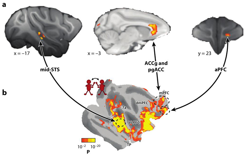
Activity in a network of areas spanning the superior temporal sulcus, dorsomedial frontal cortex, and anterior cingulate cortex is concerned with how nonhuman primates negotiate the social worlds in which they live. Central aspects of these circuits are retained in humans. Activity in these areas codes for primates' interactions with one another, their attempts to find out about one another, and their attempts to prevent others from finding out too much about themselves. Moreover, important features of the social world, such as dominance status, cooperation, and competition, modulate activity in these areas. We consider the degree to which activity in these regions is simply encoding an individual's own actions and choices or whether this activity is especially and specifically concerned with social cognition. Recent advances in comparative anatomy and computational modeling may help us to gain deeper insights into the nature and boundaries of primate social cognition.

Keywords: cingulate cortex; dominance; dorsomedial prefrontal cortex; social network; superior temporal sulcus.


Figures

Figure 1
Figure 1. Brain networks of social cognition in macaques.
Complementary whole-brain neuroimaging approaches identify mid-STS, ACCg, pgACC, and aPFC. (a) These regions are related to the size of the social group a macaque lives in (Sallet et al. 2011), and (b) they are amongst the regions more active when macaques observe conspecifics interact (Sliwa & Freiwald 2017). Panel a and b are reprinted and modified from Sallet and colleagues (2011) and from Sliwa & Freiwald (2017), respectively. Both panels reprinted with permission from AAAS. Abbreviations: ACCg, anterior cingulate gyrus; aPFC, anterior prefrontal cortex; pgACC, perigenual anterior cingulate cortex; STS, superior temporal sulcus.
Figure 2
Figure 2. Processing of facial expressions and gaze direction in STS (Morin et al. 2015).
(a) Using fMRI, patches sensitive to faces (red) are found in macaque STS when monkeys observe stimuli from different visual categories. Neurons recorded from these face-selective regions and adjacent cortex (arrow) are sensitive not only to the identity of the observed monkey but also to their gaze direction and facial expression. Example stimuli are shown in panel b, and the distribution of neuron selectivities is shown in panel c. (d) Macaque mid-STS (left) and human TPJ (right) resemble one another in the way in which their activity levels are coupled with those in other brain regions; they share aspects of their connectional fingerprints (Mars et al. 2013). Although macaque mid-STS may have the connectional fingerprint that best matches human TPJs, the areas are not identical (Mars et al. 2013). Macaque STS also resembles another human brain area in anterior STS, suggesting all three areas—human anterior STS, human TPJ, and macaque mid-STS— have some relationship. Both human TPJ and anterior STS are active during social cognitive tasks such as ToM (Deen et al. 2015, Mars et al. 2013). Panels a, b, and c adapted from Morin and colleagues (2015) by permission from Oxford University Press. Panel d adapted from Mars and colleagues (2013). Abbreviations: STS, superior temporal sulcus; ToM, theory of mind; TPJ, temporoparietal junction.
Figure 3
Figure 3. Locations of mPFC responses during social and nonsocial tasks in humans (top) and macaques (bottom).
(a) Studies that have identified responses in mPFC during fMRI in humans and the responses’ anatomical locations are represented on an MRI scan of the medial surface. Red triangles show anterior cingulate sulcus BOLD response during nonsocial decision making. Circles show locations of BOLD response that tracks social information processing. (b) Studies that have recorded from single neurons in mPFC in macaques and the neurons’ anatomical locations represented on an MRI scan of the medial surface. Red diamond shows recording site for ACCs that contains neurons that respond during nonsocial reward decision-making tasks. Rectangles show locations of neurons that code for social information. Images on the right taken from Apps and colleagues (2016) and adapted from Rushworth and colleagues (2004). Abbreviations: ACCg, anterior cingulate cortex gyrus; ACCs, anterior cingulate sulcus; BOLD, blood-oxygen-level-dependent; mPFC, medial prefrontal cortex; SFG, superior frontal gyrus.
Figure 4
Figure 4. Self- and other-values in social interactions.
(a) Dal Monte and colleagues (2017) measured social attention using eye tracking when two macaques were seated directly across from each other. Administration of OTNAL led to increased fixation on the other’s face and in particular the eyes (left, heat map of fixations under OTNAL compared to a SAL control; right, frequency of eye fixation per condition). (b) Kumaran and colleagues (2016) studied learning about one’s own (yellow) or a close friend’s (blue) place in a dominance hierarchy. For hierarchies including oneself only, the updating of hierarchy position during learning was specifically correlated with pgACC activity. (c) Self-value and other-value can be learned by tracking how well actions are performed. Self-other-mergence occurs when the performance is attributed to the inappropriate agent and people increase confidence in their own abilities after a partner has performed well (Wittmann et al. 2016b). (d) Using logistic General Linear Models (GLMs), self-performance and other-performance were used to predict self-value and other-value as measured by independent ratings. Each data point shows average beta weights (+/− SEMs) for both predictors. GLMs were performed separately for cooperation and competition conditions. Self-value and other-value were largely governed by appropriate value assignment, but self-other-mergence became apparent when considering the cooperative or competitive context. Self-value was significantly more positively influenced by other-performance in cooperation compared to competition. The analogous effect was observed for other-value. (*, p< 0.005) (e) In the same study, pgACC and dmPFC showed distinct patterns of activity. While pgACC tracked the success of one’s own recent actions, dmPFC activity reflected the impact (as shown in panel d) that interacting with others had on self- and other-values. Panel a adapted from Dal Monte and colleagues (2017). Panel b adapted from Kumaran and colleagues (2016; DOI: 10.1016/j.neuron.2016.10.052). Panel d and e adapted from Wittmann and colleagues (2016; DOI: 10.1016/j.neuron.2016.06.022). Abbreviations: dmPFC, dorsomedial prefrontal cortex; GLM, general linear model; NAL, naloxone; OT, oxytocin; OTNAL, oxytocin combined with naloxone; pgACC, perigenual anterior cingulate cortex; SAL, saline, SEM, standard error of the mean.

Similar articles

Cited by

References

    1. Akaishi R, Kolling N, Brown JW, Rushworth M. Neural mechanisms of credit assignment in a multicue environment. J Neurosci. 2016;36:1096–112. - PMC - PubMed
    1. Amodio DM, Frith CD. Meeting of minds: the medial frontal cortex and social cognition. Nat Rev Neurosci. 2006;7:268–77. - PubMed
    1. Apps MAJ, Green R, Ramnani N. Reinforcement learning signals in the anterior cingulate cortex code for others’ false beliefs. NeuroImage. 2013a;64:1–9. - PubMed
    1. Apps MAJ, Lesage E, Ramnani N. Vicarious reinforcement learning signals when instructing others. J Neurosci. 2015;35:2904–13. - PMC - PubMed
    1. Apps MAJ, Lockwood PL, Balsters JH. The role of the midcingulate cortex in monitoring others’ decisions. Front Neurosci. 2013b;7:251. - PMC - PubMed

Publication types

LinkOut - more resources