Review
. 2018 Mar 21;8(2):14.
doi: 10.3390/biom8020014.

p53-Mediated Molecular Control of Autophagy in Tumor Cells

Affiliations
Review

p53-Mediated Molecular Control of Autophagy in Tumor Cells

Maria Mrakovcic et al. Biomolecules. .

Abstract

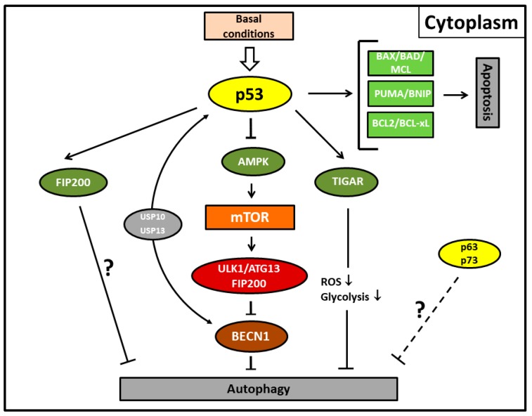
Autophagy is an indispensable mechanism of the eukaryotic cell, facilitating the removal and renewal of cellular components and thereby balancing the cell's energy consumption and homeostasis. Deregulation of autophagy is now regarded as one of the characteristic key features contributing to the development of tumors. In recent years, the suppression of autophagy in combination with chemotherapeutic treatment has been approached as a novel therapy in cancer treatment. However, depending on the type of cancer and context, interference with the autophagic machinery can either promote or disrupt tumorigenesis. Therefore, disclosure of the major signaling pathways that regulate autophagy and control tumorigenesis is crucial. To date, several tumor suppressor proteins and oncogenes have emerged as eminent regulators of autophagy whose depletion or mutation favor tumor formation. The mammalian cell "janitor" p53 belongs to one of these tumor suppressors that are most commonly mutated in human tumors. Experimental evidence over the last decade convincingly reports that p53 can act as either an activator or an inhibitor of autophagy depending on its subcellular localization and its mode of action. This finding gains particular significance as p53 deficiency or mutant variants of p53 that accumulate in the cytoplasm of tumor cells enable activation of autophagy. Accordingly, we recently identified p53 as a molecular hub that regulates autophagy and apoptosis in histone deacetylase inhibitor-treated uterine sarcoma cells. In light of this novel experimental evidence, in this review, we focus on p53 signaling as a mediator of the autophagic pathway in tumor cells.

Keywords: autophagy; histone deacetylase inhibitor; mTOR; p53; suberoylanilide hydroxamic acid; tumor.


Conflict of interest statement

The authors declare no conflict of interest.

Figures

Figure 1
Figure 1
Autophagy, apoptosis, and cell cycle arrest mediated by the activity of nuclear p53 protein (p53) in its function as a transcription factor in stress conditions. p53 primarily induces the canonical pathway of autophagy by transcriptionally upregulating tuberous sclerosis complex 2 (TSC2) (or phosphatase and tensin homolog (PTEN); not shown) and AMP-activated protein kinase (AMPK) (or the AMPK activating sestrins; not shown), thereby suppressing mammalian target of rapamycin (mTOR) and the unc-51-like autophagy activating kinase 1 (ULK1) complex including the autophagy-related protein13 (ATG13) and the focal adhesion kinase interacting protein of 200 kDa (FIP200) further downstream. For ULK1-mediated autophagosome formation or suppression, mTOR interacts further downstream with Beclin-1 (BECN1). Alternatively, damage-regulated autophagy modulator (DRAM), death-associated protein kinase (DAPK), or autophagy-related protein5 (ATG5) upregulation by the p53 family members p63 and p73 are also able to initiate autophagy. DAPK executes autophagy by phosphorylating Beclin-1 or inhibiting LC3-interacting MAP1B protein (MAP1B-LC3). DRAM and p63/p73 can also induce apoptosis. Pro-apoptotic proteins of the Bcl-2 family such as BCL2, BCL-xL, BAX, BAD, MCL, PUMA, and BNIP and the alternate reading frame protein product of the CDKN2A locus (p14ARF) can further induce autophagy via reversal of Beclin-1 inhibition. Fork symbols: inhibition by indicated proteins. Arrow lines: upregulation or activation by indicated proteins. Double arrow: major pathway activity. p53-mediated upregulation of the cyclin-dependent kinase inhibitor 1 (p21) enforces cell cycle arrest.
Figure 2
Figure 2
Autophagy and apoptosis mediated by the cytoplasmic activity of p53 protein under basal conditions. Cytoplasmic p53 protein inhibits autophagic cell death by inducing Beclin-1 (BECN1) degradation via the ubiquitin-specific peptidases USP10 and USP13 and/or inhibiting the AMPK-mTOR-ULK1 signaling pathway. It is unclear whether the canonical pathway is mediated by direct p53/FIP200 interaction or whether this represents an extra pathway. TP53-induced glycolysis and apoptosis regulator (TIGAR) inhibits autophagy by downregulation of glycolysis and suppression of reactive oxygen species (ROS) formation. p63/p73 possibly also possess transcription-independent inhibitory functions for autophagy (dashed line). Fork symbols: inhibition by indicated proteins. Arrow lines: activation by indicated proteins; downward arrow, downregulation. For abbreviations, see Figure 1.
Figure 3
Figure 3
Illustration showing presumed mechanisms mediating suberoylanilide hydroxamic acid (SAHA)-induced autophagy. (A) Acetylated cytoplasmic p53 protein that escapes deacetylating activity by applying HDACi (histone deacetylase inhibitor) preferentially induces apoptotic cell death by direct interaction with the Bcl-2 family of pro-apoptotic proteins. Concurrently, cytoplasmic p53 protein inhibits autophagic cell death by inducing Beclin-1 degradation via the ubiquitin-specific peptidases USP10 and USP13 and/or inhibiting the AMPK-mTOR-ULK1 signaling pathway. It is unclear whether the canonical pathway is mediated by direct p53/FIP200 interaction or whether this represents an extra pathway. TP53-induced glycolysis and apoptosis regulator (TIGAR) inhibits autophagy by downregulation of glycolysis and a suppression of ROS formation. The p53 family members p63/p73 possibly also possess transcription-independent inhibitory functions for autophagy (dashed line). (B) Mutant p53 (p53Mut) protein reverses the situation and predominantly activates autophagy due to its inability to inhibit autophagy or stimulate apoptosis. Fork symbols: inhibition by indicated proteins. Arrow lines: activation or interaction with indicated proteins. Double arrow: major pathway activity. For abbreviations, see Figure 1.

References

    1. Mizushima N., Noda T., Yoshimori T., Tanaka Y., Ishii T., George M.D., Klionsky D.J., Ohsumi M., Ohsumi Y. A protein conjugation system essential for autophagy. Nature. 1998;395:395–398. doi: 10.1038/26506. - DOI - PubMed
    1. Yang Z., Klionsky D.J. Mammalian autophagy: Core molecular machinery and signaling regulation. Curr. Opin. Cell Biol. 2010;22:124–131. doi: 10.1016/j.ceb.2009.11.014. - DOI - PMC - PubMed
    1. Mizushima N., Komatsu M. Autophagy: Renovation of cells and tissues. Cell. 2011;147:728–741. doi: 10.1016/j.cell.2011.10.026. - DOI - PubMed
    1. Li W.W., Li J., Bao J. Microautophagy: lesser-known self-eating. Cell. Mol. Life Sci. 2012;69:1125–1136. doi: 10.1007/s00018-011-0865-5. - DOI - PMC - PubMed
    1. Kaushik S., Cuervo A.M. Chaperone-mediated autophagy: A unique way to enter the lysosome world. Trends Cell Biol. 2012;22:305–309. doi: 10.1016/j.tcb.2012.05.006. - DOI - PMC - PubMed

Publication types

Substances