Review
. 2018 Mar 21;9(4):174.
doi: 10.3390/genes9040174.

The Role of microRNAs in Alzheimer's Disease and Their Therapeutic Potentials

Affiliations
Review

The Role of microRNAs in Alzheimer's Disease and Their Therapeutic Potentials

Munvar Miya Shaik et al. Genes (Basel). .

Abstract

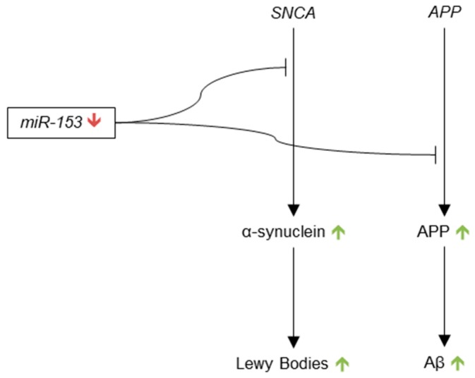
MicroRNAs (miRNAs) are short, endogenous, non-coding RNAs that post-transcriptionally regulate gene expression by base pairing with mRNA targets. Altered miRNA expression profiles have been observed in several diseases, including neurodegeneration. Multiple studies have reported altered expressions of miRNAs in the brains of individuals with Alzheimer's disease (AD) as compared to those of healthy elderly adults. Some of the miRNAs found to be dysregulated in AD have been reported to correlate with neuropathological changes, including plaque and tangle accumulation, as well as altered expressions of species that are known to be involved in AD pathology. To examine the potentially pathogenic functions of several dysregulated miRNAs in AD, we review the current literature with a focus on the activities of ten miRNAs in biological pathways involved in AD pathogenesis. Comprehensive understandings of the expression profiles and activities of these miRNAs will illuminate their roles as potential therapeutic targets in AD brain and may lead to the discovery of breakthrough treatment strategies for AD.

Keywords: Alzheimer’s disease; BACE1 inhibitors; miRNAs; γ-secretase inhibitors.


Conflict of interest statement

The authors declare no conflict of interest.

Figures

Figure 1
Figure 1
Regulation by miR-9. miR-9 inhibits the translation of β-site amyloid 1102 precursor protein cleaving enzyme 1 (BACE1), transforming growth factor, β-induced (TGFBI), tripartite motif-containing 2 (TRIM2), silent mating type information regulation 2 homolog 1 (SIRT1), and calcium/calmodulin-dependent protein kinase kinase 2 (CAMKK2). The debatable level of miR-9 expression in Alzheimer’s disease (AD) brain is indicated by the yellow tilde symbol (~), as are the hypothetical expression levels of the proteins regulated by miR-9 and their cellular effects. ROS: reactive oxygen species.
Figure 2
Figure 2
Regulation by microRNA (miR)-124. miR-124 inhibits the translation of BACE1 and Polypyrimidine Tract Binding Protein 1 (PTBP1). The down-regulation of miR-124 in AD brain is indicated by the red arrow. The hypothetical up-regulations of the target proteins and their cellular effects are denoted by the green arrows.
Figure 3
Figure 3
Regulation by miR-181. miR-181 inhibits the translation of Serine Palmitoyltransferase Long Chain Base Subunit 1 (SPTLC1), tripartite motif-containing 2 (TRIM2), silent mating type information regulation 2 homolog 1 (SIRT1), BTB Domain Containing 3 (BTBD3), high-mobility group protein 1 (HMGB1), B-cell lymphoma 2 (Bcl2), Nicotinamide phosphoribosyltransferase (NAMPT), methyl CpG binding protein 2 (MeCP2), and X-linked inhibitor of apoptosis (XIAP). The decreased levels of miR-181 in AD brain are denoted by the red arrow. The green arrows denote the hypothetical upregulation of the targeted proteins and their cellular effects.
Figure 4
Figure 4
Regulation by miR-29. miR-29 inhibits the translation of BACE1, several Bcl-2 Homology 3 (BH3)-only family proteins (Bim, Bmf, Hrk, Puma, and N-bak), SPTLC2 and neuron navigator 3 (NAV3). The debatable levels of miR-29 expression in AD brain are indicated by the yellow tilde symbol (~), as are the hypothetical expression levels of the proteins regulated by miR-29 and their cellular effects.
Figure 5
Figure 5
Regulation by miR-34. miR-34 inhibits the translation of BCL2, SIRT1, and Tau. The green arrow indicates the increased expression levels of miR-34 in AD brain. The red arrows indicate the hypothetically decreased levels of the targeted proteins and their cellular effects.
Figure 6
Figure 6
Regulation by miR-107. miR-107 inhibits the translation of BACE1, GRN, CF1, CDK5R1, and ADAM10. The red arrow indicates the downregulation of miR-107 observed in AD brain. The green arrows indicated the hypothetically increased levels of the proteins targeted by miR-107, as well as their cellular effects.
Figure 7
Figure 7
Regulation of miR-146. miR-146 expression is induced by multiple inflammatory species (Tumor necrosis factor (TNF)-α (TNFα), nuclear factor kappa-B (NF-κB), H2O2, interleukin (IL)-1β (IL-1β), and reactive oxygen species (ROS)), as well as Aβ. The yellow tilde (~) indicates the debated levels of miR-146 in AD brain.
Figure 8
Figure 8
Regulation by miR-101. miR-101 inhibits the translation of APP. The red arrow indicates the significant downregulation of miR-101 expression observed in AD brain. The green arrows indicated the hypothetically increased levels of APP and Aβ resulting from the downregulation of miR-101.
Figure 9
Figure 9
Regulation by miR-195. miR-195 inhibits the translation of BACE1. The yellow tildes (~) indicate the lack of consensus on the expression levels of miR-195 in AD brain, as well as the hypothetically unclear effects miR-195 on the levels of BACE1 and Aβ.
Figure 10
Figure 10
Regulation by miR-153. miR-153 inhibits the translation of α-synuclein and amyloid precursor protein (APP). The red arrow indicates that miR-153 expression is decreased in AD brain. The green arrows denote the hypothetically increased levels of α-synuclein, Lewy bodies, APP, and Aβ resulting from miR-153 downregulation.

References

    1. Jellinger K.A. Clinicopathological analysis of dementia disorders in the elderly—An update. J. Alzheimers Dis. 2006;9:61–70. doi: 10.3233/JAD-2006-9S308. - DOI - PubMed
    1. Jellinger K., Danielczyk W., Fischer P., Gabriel E. Clinicopathological analysis of dementia disorders in the elderly. J. Neurol. Sci. 1990;95:239–258. doi: 10.1016/0022-510X(90)90072-U. - DOI - PubMed
    1. McKhann G.M., Knopman D.S., Chertkow H., Hyman B.T., Jack C.R., Kawas C.H., Klunk W.E., Koroshetz W.J., Manly J.J., Mayeux R., et al. The diagnosis of dementia due to Alzheimer’s disease: Recommendations from the national institute on aging-alzheimer’s association workgroups on diagnostic guidelines for Alzheimer’s disease. Alzheimers Dement. 2011;7:263–269. doi: 10.1016/j.jalz.2011.03.005. - DOI - PMC - PubMed
    1. Jack C.R., Albert M.S., Knopman D.S., McKhann G.M., Sperling R.A., Carrillo M.C., Thies B., Phelps C.H. Introduction to the recommendations from the national institute on aging-Alzheimer’s association workgroups on diagnostic guidelines for alzheimer’s disease. Alzheimers Dement. 2011;7:257–262. doi: 10.1016/j.jalz.2011.03.004. - DOI - PMC - PubMed
    1. Sperling R.A., Aisen P.S., Beckett L.A., Bennett D.A., Craft S., Fagan A.M., Iwatsubo T., Jack C.R., Kaye J., Montine T.J., et al. Toward defining the preclinical stages of alzheimer’s disease: Recommendations from the national institute on aging-alzheimer’s association workgroups on diagnostic guidelines for alzheimer’s disease. Alzheimers Dement. 2011;7:280–292. doi: 10.1016/j.jalz.2011.03.003. - DOI - PMC - PubMed