. 2018 Mar 21;14(3):e1007029.
doi: 10.1371/journal.pgen.1007029. eCollection 2018 Mar.

Cdk1 phosphorylation of Esp1/Separase functions with PP2A and Slk19 to regulate pericentric Cohesin and anaphase onset

Affiliations

Cdk1 phosphorylation of Esp1/Separase functions with PP2A and Slk19 to regulate pericentric Cohesin and anaphase onset

Noel Lianga et al. PLoS Genet. .

Abstract

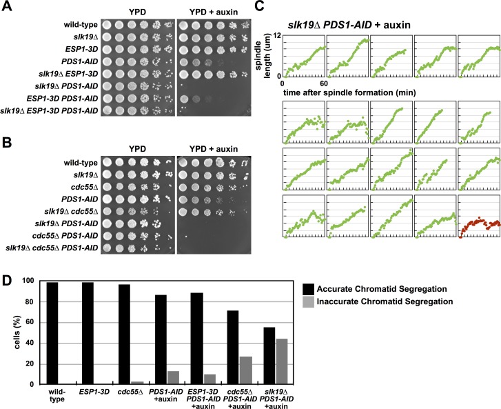
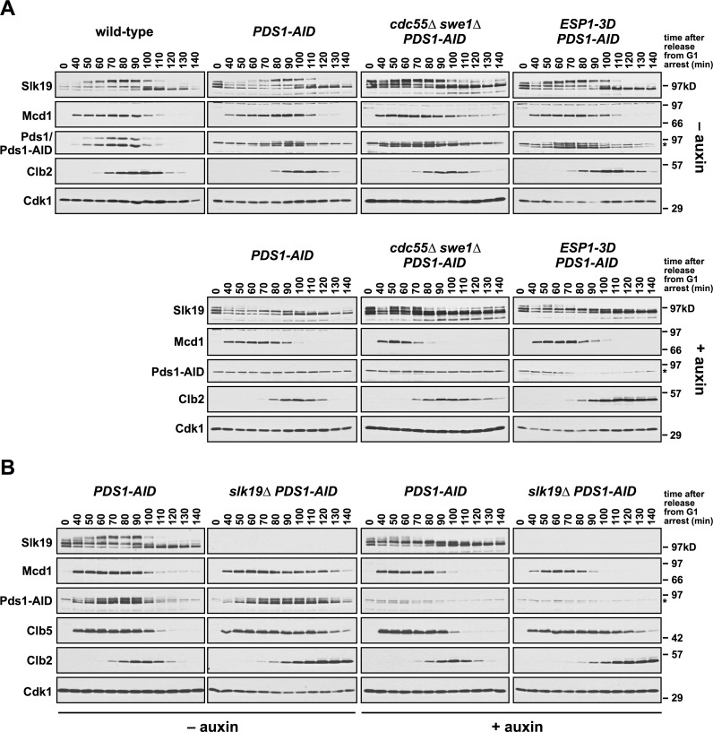
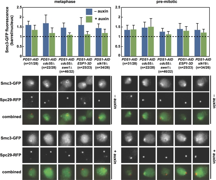
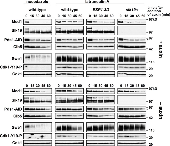
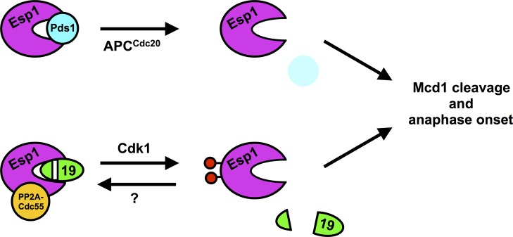
Anaphase onset is an irreversible cell cycle transition that is triggered by the activation of the protease Separase. Separase cleaves the Mcd1 (also known as Scc1) subunit of Cohesin, a complex of proteins that physically links sister chromatids, triggering sister chromatid separation. Separase is regulated by the degradation of the anaphase inhibitor Securin which liberates Separase from inhibitory Securin/Separase complexes. In many organisms, Securin is not essential suggesting that Separase is regulated by additional mechanisms. In this work, we show that in budding yeast Cdk1 activates Separase (Esp1 in yeast) through phosphorylation to trigger anaphase onset. Esp1 activation is opposed by protein phosphatase 2A associated with its regulatory subunit Cdc55 (PP2ACdc55) and the spindle protein Slk19. Premature anaphase spindle elongation occurs when Securin (Pds1 in yeast) is inducibly degraded in cells that also contain phospho-mimetic mutations in ESP1, or deletion of CDC55 or SLK19. This striking phenotype is accompanied by advanced degradation of Mcd1, disruption of pericentric Cohesin organization and chromosome mis-segregation. Our findings suggest that PP2ACdc55 and Slk19 function redundantly with Pds1 to inhibit Esp1 within pericentric chromatin, and both Pds1 degradation and Cdk1-dependent phosphorylation of Esp1 act together to trigger anaphase onset.


Conflict of interest statement

The authors have declared that no competing interests exist.

Figures

Fig 1
Fig 1. Cdk1Clb2 phosphorylates Esp1.
(A) Esp1 is phosphorylated in vivo. ESP1, ESP1-myc13 and pGAL-SWE1 ESP1-13myc cells were grown in YEP + raffinose, arrested in mitosis with nocodazole and switched to YEP + galactose media to induce expression of Swe1. After 1 h, cells were washed in medium lacking phosphate, and grown for 30 min in the presence of [32P]orthophosphate. Esp1-13myc was immunoprecipitated with 9E10 antibody, run on a polyacrylamide gel and exposed to a phosphorimager screen or immunoblotted. (B) Esp1 contains six minimal Cdk1 consensus sites (S/TP): S13 and T16 (termed N-terminal), T1014, S1027 and T1034 (termed central) and S1280 (termed C-terminal). (C) Mutating Esp1 central residues prevents Esp1 phosphorylation in vivo. Left panels, ESP1, esp1-2A, esp1-3A, esp1-1A, esp1-3A+2A, esp1-2A+1A, esp1-3A+1A and esp1-2A+3A+1A cells were grown in YEP + dextrose, arrested in mitosis with nocodazole and labeled with [32P]orthophosphate as described in (A). Right panels, wild-type, cdc55Δ and rts1Δ cells were grown in YEP + dextrose, arrested in G1 with α-factor and released into the cell cycle in the presence of nocodazole. After 90 min cells were labeled with [32P]orthophosphate as described in (A). Esp1 was immunoprecipitated with anti-Esp1 antibody, run on a polyacrylamide gel and exposed to a phosphorimager screen or immunoblotted. (D) Wild-type and esp1-3A cells were grown to log phase, arrested in G1 with α-factor, and released into the cell cycle (t = 0). α-factor was re-added at t = 60 min to arrest cells in the following G1. Samples were taken for immunoblotting at the indicated timepoints, and run on a polyacrylamide gel containing Phos-tag reagent (top panel), or a standard polyacrylamide gel (bottom panels) and immunoblotted with the indicated antibodies. Note that running and transferring of Phos-tag polyacrylamide gels is inconsistent and cell cycle-dependent changes in protein abundance observed in these panels may not accurately reflect changes in protein abundance. The Esp1 panels from the standard polyacrylamide gel more accurately reflect cell cycle changes in Esp1 abundance. (E) Cdk1Clb2 phosphorylates the central region of Esp1 in vitro. Esp1 was immunoprecipitated from the strains in (C) growing asynchronously, incubated with γ-[32P]ATP and purified Cdk1Clb2-CBP, washed, run on a polyacrylamide gel, and exposed to a phosphorimager screen or immunoblotted with anti-Esp1 antibody. (F) PP2ACdc55 dephosphorylates Esp1 in vitro. Esp1 was immunoprecipitated from wild-type cells and phosphorylated with purified Cdk1Clb2-CBP and γ-[32P]ATP while immobilized on IgG-coupled magnetic beads. The beads were washed and incubated for the indicated times at room temperature with no addition (yellow lines), TAP-purified PP2ACdc55 (blue lines), or PP2ACdc55 and okadaic acid (OA) (red lines). The three reactions share a t = 0 sample that was taken before the additions. The dephosphorylation of Esp1 was quantified on a phosphorimager and the extent of dephosphorylation relative to t = 0 (average ± SEM) was graphed. The experiment shown is representative of one of three repeats.
Fig 2
Fig 2. Pds1 depletion causes immediate spindle elongation in a phospho-mimetic Esp1 mutant.
(A) Pds1 depletion causes synthetic sickness in ESP1-3D cells. Eight-fold serial dilutions of the indicated strains were spotted onto the indicated plates and grown at 25°C. (B) The ESP1-3D allele is semi-dominant in diploids. Ten-fold serial dilutions of the indicated diploids were grown in YEP + dextrose media, spotted onto the indicated plates and grown at 25°C. (C) The ESP1-3D allele is semi-dominant when complemented by a plasmid borne ESP1 (ESP1-CEN-HIS3). Ten-fold serial dilutions of the indicated strains were grown in media lacking histidine, spotted onto the indicated plates and grown at 25°C. (D) Mitotic spindle morphology of individual ESP1-3D cells depleted of Pds1. PDS1-AID SPC42-eGFP and ESP1-3D PDS1-AID SPC42-eGFP cells were grown at 25°C to log phase and arrested in G1 with α-factor. 30 min before α-factor release +/- auxin was added. Cells were released at t = 0 and at t = 25 min cells were plated onto YPD live microscopy pads +/- auxin and Spc42-eGFP was imaged every minute. Each strain was imaged at least two times in each condition. Cells that undergo normal metaphase spindle formation are shown in black. Cells that undergo immediate spindle elongation upon spindle formation are shown in green. Cells that exhibit failed or no anaphase spindle elongation are shown in red. See S3 Fig for cell traces of experiments done in the absence of auxin and Fig 3 for tabulation of all imaging data. The scoring metric is described in the text and in the material and methods.
Fig 3
Fig 3. Spindle characteristics during mitosis.
Cells with “normal metaphase spindle formation” do not elongate their spindle more than 2 μm in the first 10 minutes after SPB separation and more than 2.5 μm in the first 15 minutes after SPB separation. Cells whose spindles elongate more than 2 and 2.5 μm in these time-intervals display “immediate spindle elongation.” Cells with “failed anaphase” don’t elongate their spindles to 6 μm in the 60 minutes after SPB separation. A small number of cells (15% for cells treated with auxin, and 11% for untreated cells) produce conflicting scores using these two rules (i.e., immediate/normal or normal/immediate in the 10/15 minute intervals), and we categorized these cells manually (see Materials and Methods for details).
Fig 4
Fig 4. Pds1 depletion in cdc55Δ cells induce premature anaphase onset and chromosome mis-segregation.
(A) cdc55Δ PDS1-AID cells are inviable on media containing auxin. Eight-fold serial dilutions of the indicated strains were spotted onto the indicated plates and grown at 25°C. (B) Mitotic spindle morphology of individual cdc55Δ cells depleted of Pds1. cdc55Δ PDS1-AID SPC42-eGFP and swe1Δ cdc55Δ PDS1-AID SPC42-eGFP cells were imaged as in Fig 2D. See S3 Fig for cell traces of experiments done in the absence of auxin, and Fig 3 for tabulation of all imaging data. (C) Pds1 depletion in cdc55Δ cells cause premature sister chromatid separation. PDS1-AID, cdc55Δ PDS1-AID and ESP1-3D PDS1-AID cells containing ura3::240lacO pCUP1-eGFP-lacI SPC42-mCherry were imaged as in Fig 2D. Data from individual experiments were combined for subsequent analysis (PDS1-AID–auxin [n = 17], PDS1-AID + auxin [n = 26], cdc55Δ PDS1-AID + auxin [n = 14], and ESP1-3D PDS1-AID [n = 19]). The length of time between spindle formation and sister chromatid separation was determined for each imaged cell. Cells in which sister chromatids segregated to the same pole during anaphase were characterized as failed and analyzed separately. If sister chromatid separation preceded spindle formation, this length of time was defined as 0. All measured times are displayed with the population average (± SEM). The time between spindle formation and sister separation in cdc55Δ cells is significantly different from auxin-treated and untreated PDS1-AID cells (Student’s t-tests; p < 0.05), and in auxin-treated PDS1-AID cells is significantly different from untreated PDS1-AID cells (Student’s t-test; p < 0.05). (D) An example of one cdc55Δ PDS1-AID cell imaged in (C). Inter-spindle pole body (spindle length; red squares) and inter-sister chromatid (green diamonds) distance is graphed, and in this cell, sister chromatid separation preceded spindle formation, and both sisters segregated to the same pole. (E) Pds1 depletion in cdc55Δ cells cause failed sister chromatid segregation. Segregation of sister chromatids was monitored in the cells imaged in (C). Cells in which sister chromatids segregated to the same pole during anaphase were characterized as failed. Cells in which sister chromatids segregated to the separate poles, regardless of timing were characterized as accurate. PDS1-AID–auxin (n = 17), PDS1-AID + auxin (n = 36), cdc55Δ PDS1-AID + auxin (n = 27).
Fig 5
Fig 5. mcd1-10A and esp1-3A do not suppress cdc55Δ phenotypes.
(A) esp1-3A, mcd1-10A and esp1-3A mcd1-10A do not rescue cdc55Δ cells depleted of Pds1. Ten-fold serial dilutions of the indicated strains were spotted onto the indicated plates and grown at 25°C. (B) Cdc14 is released at a spindle length of approximately 2 μm in swe1Δ cdc55Δ cells regardless of Pds1 depletion. swe1Δ cdc55Δ PDS1-AID CDC14-eGFP SPC42-mCherry cells were grown at 25˚C to log phase and arrested in G1 with α-factor. 30 min before α-factor release +/- auxin was added. Cells were released at t = 0 and at t = 90 min samples were fixed for microscopy. The distance between spindle pole bodies was measured in each cell. Each cell was categorized as pre-mitotic (a single Spc42-mCherry focus), short spindle (Spc42-mCherry foci separated by < 2μm) or long spindle (Spc42-mCherry foci separated by > 2μm). In each cell Cdc14 was characterized as nucleolar or released qualitatively. The proportion of cells displaying nucleolar and released Cdc14 at each cell cycle stage relative to spindle length was calculated. swe1Δ cdc55Δ PDS1-AID–auxin (n = 88), swe1Δ cdc55Δ PDS1-AID + auxin (n = 102). All raw images are shown in S4A Fig.
Fig 6
Fig 6. Pds1 depletion in slk19Δ cells induce premature anaphase onset and chromosome mis-segregation.
(A) slk19Δ PDS1-AID cells are inviable in media containing auxin, and slk19Δ does not interact genetically with ESP1-3D. Eight-fold serial dilutions of the indicated strains were spotted onto the indicated plates and grown at 25°C. (B) slk19Δ does not interact genetically with cdc55Δ. Eight-fold serial dilutions of the indicated strains were spotted onto the indicated plates and grown at 25°C. (C) Mitotic spindle morphology of individual slk19Δ cells depleted of Pds1. slk19Δ PDS1-AID SPC42-eGFP cells were imaged as in Fig 2D. See S3 Fig for cell traces of experiments done in the absence of auxin, and Fig 3 for tabulation of all imaging data. (D) cdc55Δ and slk19Δ cells mis-segregate chromosomes. The indicated strains containing pCUP1-eGFP-lacI ura3::240lacO SPC42-mCherry were grown at 25°C to log phase and arrested in G1 with α-factor. 30 min before α-factor release auxin was added. Cells were released at t = 0 and at t = 120 min samples were fixed for microscopy. Telophase cells were identified using the Spc42-mCherry signal. The presence of GFP-lacI foci at either both spindle poles or at a single spindle pole was scored (n = 100 for each strain).
Fig 7
Fig 7. Pds1 depletion in cdc55Δ swe1Δ and slk19Δ cells induce premature Slk19 cleavage and Mcd1 proteolysis.
(A) The indicated strains were grown to log phase, arrested in G1 with α-factor, and 30 min before α-factor release +/- auxin was added to the cultures. Cells were released from the arrest (t = 0) at 25°C into media containing +/- auxin. α-factor was added at t = 80 min to arrest cells in the following G1. Samples were harvested for immunoblotting at the indicated timepoints, run on a polyacrylamide gel, and immunoblotted with the indicated antibodies. Wild-type cells were grown in parallel, but not treated with auxin and are included as a comparison to untreated PDS1-AID cells. Pds1-AID migrates adjacent to a background band (indicated by an *). (B) PDS1-AID and slk19Δ PDS1-AID cells were grown as in part (A). Samples were harvested for immunoblotting at the indicated timepoints, run on a polyacrylamide gel, and immunoblotted with the indicated antibodies. Pds1-AID migrates adjacent to a background band (indicated by an *).
Fig 8
Fig 8. The pericentric Cohesin barrel does not form when Pds1 is depleted from cdc55Δ, cdc55Δ swe1Δ, ESP1-3D and slk19Δ cells.
The indicated strains containing SMC3-GFP SPC29-RFP were imaged as in Fig 2D. Maximal Cohesin fluorescence between SPBs was measured in cells with spindles shorter than 2 μm (metaphase, left), and in cells with a single SPB (pre-mitotic, right). The ratio of barrel/nuclear fluorescence is plotted (average ± SD). A value of 1 is the background nuclear fluorescence. There are significant differences between all untreated and auxin-treated strains (p < 0.05; Student’s t-tests).
Fig 9
Fig 9. ESP1-3D and slk19Δ bypass a Pds1-independent arrest.
The indicated strains were grown to log phase, arrested in G1 with α-factor, and released from the arrest into media containing nocodazole or latrunculin A at 25°C. After 2 h (t = 0), +/- auxin was added to the cultures. Samples were harvested for immunoblotting at the indicated timepoints, run on a polyacrylamide gel, and immunoblotted with the indicated antibodies. cdc55Δ was not analyzed in this experiment because it is defective in both the spindle assembly and morphogenesis checkpoints, and does not arrest in nocodazole or LatA. Pds1-AID migrates adjacent to a background band (indicated by an *).
Fig 10
Fig 10. Proposed model for redundant inhibition of Esp1 by Pds1 and Slk19.
Our data support a model in which both Pds1 and Slk19 (19) inhibit Esp1. Anaphase onset is triggered by both APCCdc20-mediated proteolysis of Pds1 and Cdk1-dependent activation of Esp1. Slk19 and PP2ACdc55 inhibit Esp1, and Cdk1 phosphorylation of Esp1 overcomes this inhibition. We speculate that Esp1 phosphorylation triggers the release of Slk19 from the active site of Esp1. Although PP2ACdc55 can dephosphorylate Esp1 in vitro and cdc55Δ cells increase Esp1 phosphorylation in vivo, the lack of suppression of cdc55Δ by esp1-3A suggest that PP2ACdc55 binding to Separase may be more important for Esp1 inhibition. Slk19 can inhibit Separase either as an uncleaved or cleaved (as depicted) protein.

Similar articles

Cited by

References

    1. Guacci V, Koshland D, Strunnikov A. A direct link between sister chromatid cohesion and chromosome condensation revealed through the analysis of MCD1 in S. cerevisiae. Cell. 1997;91: 47–57. - PMC - PubMed
    1. Michaelis C, Ciosk R, Nasmyth K. Cohesins: chromosomal proteins that prevent premature separation of sister chromatids. Cell. 1997;91: 35–45. - PubMed
    1. Strunnikov AV, Larionov VL, Koshland D. SMC1: an essential yeast gene encoding a putative head-rod-tail protein is required for nuclear division and defines a new ubiquitous protein family. J Cell Biol. 1993;123: 1635–1648. - PMC - PubMed
    1. Gruber S, Haering CH, Nasmyth K. Chromosomal cohesin forms a ring. Cell.2003;112: 765–777. - PubMed
    1. Haering CH, Farcas A-M, Arumugam P, Metson J, Nasmyth K. The cohesin ring concatenates sister DNA molecules. Nature. 2008;454: 297–301. doi: 10.1038/nature07098 - DOI - PubMed

Publication types

MeSH terms