

Clinical value of patient-specific three-dimensional printing of congenital heart disease: Quantitative and qualitative assessments
- PMID: 29561912
- PMCID: PMC5862481
- DOI: 10.1371/journal.pone.0194333
Clinical value of patient-specific three-dimensional printing of congenital heart disease: Quantitative and qualitative assessments
Abstract
Objective: Current diagnostic assessment tools remain suboptimal in demonstrating complex morphology of congenital heart disease (CHD). This limitation has posed several challenges in preoperative planning, communication in medical practice, and medical education. This study aims to investigate the dimensional accuracy and the clinical value of 3D printed model of CHD in the above three areas.
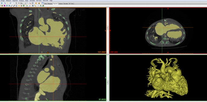
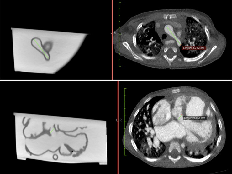
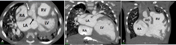
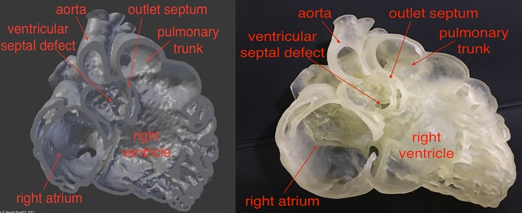
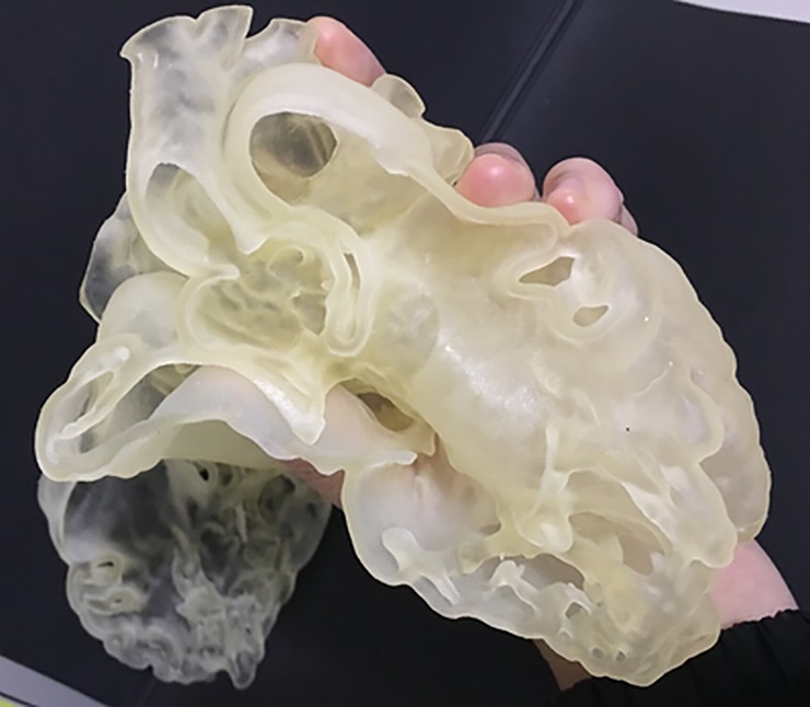
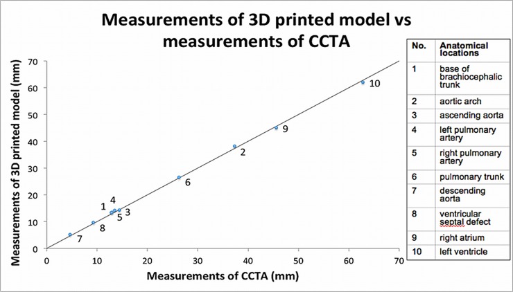
Methods: Using cardiac computed tomography angiography (CCTA) data, a patient-specific 3D model of a 20-month-old boy with double outlet right ventricle was printed in Tango Plus material. Pearson correlation coefficient was used to evaluate correlation of the quantitative measurements taken at analogous anatomical locations between the CCTA images pre- and post-3D printing. Qualitative analysis was conducted by distributing surveys to six health professionals (two radiologists, two cardiologists and two cardiac surgeons) and three medical academics to assess the clinical value of the 3D printed model in these three areas.
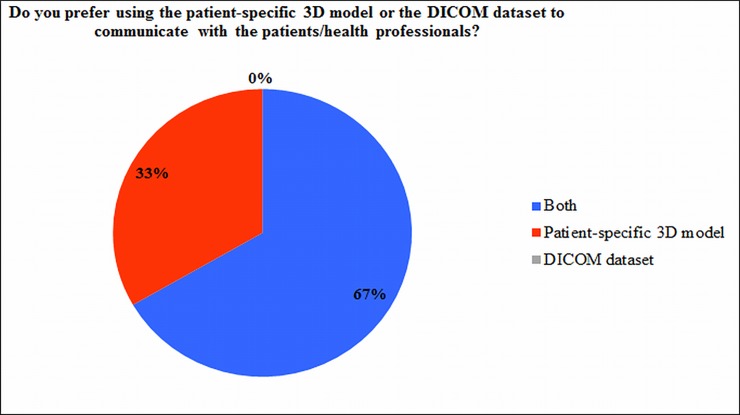
Results: Excellent correlation (r = 0.99) was noted in the measurements between CCTA and 3D printed model, with a mean difference of 0.23 mm. Four out of six health professionals found the model to be useful in facilitating preoperative planning, while all of them thought that the model would be invaluable in enhancing patient-doctor communication. All three medical academics found the model to be helpful in teaching, and thought that the students will be able to learn the pathology quicker with better understanding.
Conclusion: The complex cardiac anatomy can be accurately replicated in flexible material using 3D printing technology. 3D printed heart models could serve as an excellent tool in facilitating preoperative planning, communication in medical practice, and medical education, although further studies with inclusion of more clinical cases are needed.
Conflict of interest statement
Figures
References
-
- Australian Institute of Health and Welfare [Internet]. Australian Government; c2017. How many Australians have cardiovascular disease? [cited 2017 Feb 28]. Available from: http://www.aihw.gov.au/cardiovascular-disease/prevalence/#t5.
-
- Bhatla P, Tretter JT, Ludomirsky A, Argilla M, Latson LA Jr, S, et al. Utility and scope of rapid prototyping in patients with complex muscular ventricular septal defects or double-outlet right ventricle: Does it alter management decisions? Pediatr Cardiol. 2017;38(1):103–114. doi: 10.1007/s00246-016-1489-1 . - DOI - PubMed
-
- Schmauss D, Haeberle S, Hagl C, Sodian R. Three-dimensional printing in cardiac surgery and interventional cardiology: a single-centre experience. Eur J Cardiothorac Surg. 2015;47:1044–1052. doi: 10.1093/ejcts/ezu310 . - DOI - PubMed
-
- Valverde I, Gomez G, Gonzales A, Suarez-Mejias C, Adsuar A, Coserria JF, et al. Three-dimensional patient-specific cardiac model for surgical planning in Nikaidoh procedure. Cardiol Young. 2015;25(4):698–704. doi: 10.1017/S1047951114000742 . - DOI - PubMed
-
- Riesenkampff E, Rietdorf U, Wolf I, Schnackenburg B, Ewert P, Huebler M, et al. The practical clinical value of three-dimensional models of complex congenitally malformed hearts. J Thorac Cardiovascular Surgery. 2009;138(3):571–580. doi: 10.1016/j.jtcvs.2009.03.011 . - DOI - PubMed
Publication types
MeSH terms
LinkOut - more resources
Full Text Sources
Other Literature Sources
Medical
