Review
. 2018 Mar 18;7(1):25.
doi: 10.3390/antibiotics7010025.

Novel Aspects of Polynucleotide Phosphorylase Function in Streptomyces

Affiliations
Review

Novel Aspects of Polynucleotide Phosphorylase Function in Streptomyces

George H Jones. Antibiotics (Basel). .

Abstract

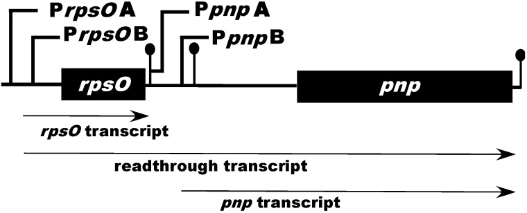
Polynucleotide phosphorylase (PNPase) is a 3'-5'-exoribnuclease that is found in most bacteria and in some eukaryotic organelles. The enzyme plays a key role in RNA decay in these systems. PNPase structure and function have been studied extensively in Escherichiacoli, but there are several important aspects of PNPase function in Streptomyces that differ from what is observed in E. coli and other bacterial genera. This review highlights several of those differences: (1) the organization and expression of the PNPase gene in Streptomyces; (2) the possible function of PNPase as an RNA 3'-polyribonucleotide polymerase in Streptomyces; (3) the function of PNPase as both an exoribonuclease and as an RNA 3'-polyribonucleotide polymerase in Streptomyces; (4) the function of (p)ppGpp as a PNPase effector in Streptomyces. The review concludes with a consideration of a number of unanswered questions regarding the function of Streptomyces PNPase, which can be examined experimentally.

Keywords: (p)ppGpp; RNA decay; Streptomyces; antibiotic; polyadenylation; polynucleotide phosphorylase; promoter; regulation; ribonuclease.


Conflict of interest statement

The author declares no conflicts of interest.

Figures

Figure 1
Figure 1
Schematic representation of the Streptomyces coelicolor rpsO–pnp operon. PrpsOA, B and PpnpA, B represent the upstream and intergenic promoters found in S. coelicolor, respectively. The ball-and-stick structures immediately following rpsO and pnp represent rho-independent transcription terminators. The ball-and-stick structure just upstream of pnp represents the intergenic hairpin which is cleaved by RNase III. The diagram is not drawn to scale.
Figure 2
Figure 2
(A) Growth of the S. coelicolor strains containing promoter probe constructs. Growth was measured as the increase in optical density at 450 nm. The arrows in the figure indicate the onset of the production of two of the secondary metabolites synthesized by S. coelicolor, undecylprodigiosin (red) and actinorhodin (act). (B) Catechol dioxygenase (CATO2ase) activity of mycelial extracts of S. coelicolor derivatives containing the putative rpsOA and rpsOB promoters, cloned in the promoter probe vector pIPP2 [35]. Mycelium was harvested at the indicated times, disrupted by sonication, and following centrifugation, supernatants were assayed for catechol dioxygenase, as described previously [31,35]. The catechol dioxygenase gene is the reporter in the promoter probe vector [35]. (C) CATO2ase activities of extracts of strains containing the putative pnpA and pnpB promoters. The results shown are the averages of duplicate assays from two independent experiments ± SEM. This figure is reprinted from Gene, 536, Patricia Bralley, Marcha L. Gatewood, George H. Jones, Transcription of the rpsO–pnp operon of Streptomyces coelicolor involves four temporally regulated, stress responsive promoters. 177–185, Copyright (2014), with permission from Elsevier [31].
Figure 3
Figure 3
Cold shock responses of S. coelicolor. Derivatives containing the rpsO–pnp promoter probe constructs were grown and 30 °C, and half of each culture was then shifted to 10 °C. Mycelium was harvested at the indicated times, disrupted by sonication, and following centrifugation, supernatants were assayed for promoter activity, as described [31,35]. Panel C shows the results of PNPase polymerization assays. In Panels A and B, PNPase promoter activities are expressed relative to the activity measured at 30 °C at zero time, immediately before the shift to 10 °C. The results shown are the averages of duplicate assays from two independent experiments ± SEM. In the first experiment, PNPase levels were measured in S. coelicolor containing PrpsOA and in the second, PNPase levels were measured in the derivative containing PpnpB. This figure is reprinted from Gene, 536, Patricia Bralley, Marcha L. Gatewood, George H. Jones, Transcription of the rpsO-pnp operon of Streptomyces coelicolor involves four temporally regulated, stress responsive promoters. 177–185, Copyright (2014), with permission from Elsevier [31].
Figure 4
Figure 4
Effects of nucleoside diphosphates on the phosphorolysis of the 5650 transcript. Phosphorolysis reactions were performed as described in [47], and reaction products were separated by gel electrophoresis. The top panel shows the results obtained with S. coelicolor PNPase and the bottom panel results using E. coli PNPase. Reactions were conducted in the presence of increasing concentrations of a mixture of ADP, CDP, UDP, and GDP (nucleoside diphosphates (NDPs)) as indicated. RP3 is the 5650 transcript, and RP4 represents the product obtained by complete digestion of the intergenic hairpin in RP3 by PNPase. Note that as PNPase is highly processive [48], no intermediates with mobilities between those of RP3 and RP4 were observed. Copyright © American Society for Microbiology (J. Bacteriol. 190, 2008, 98–106, DOI:10.1128/JB.00327-07) [47].
Figure 5
Figure 5
Model for the effects of NDPs on the activity S. coelicolor PNPase. The model posits that S. coelicolor PNPase (PacMan symbol) is able to phosphorolyze 5650 and other structured substrates to a limited extent in the absence of NDPs, as indicated by the dashed X. In the presence of NDPs, PNPase synthesizes unstructured 3′-tails in vivo, and these tails then provide an anchor for the enzyme, thus facilitating the digestion of structured substrates. Copyright © American Society for Microbiology (J. Bacteriol. 195, 2013, 5151–5159, DOI:10.1128/JB.00936-13) [49].
Figure 6
Figure 6
Effects of (p)ppGpp on the activity of PNPase. Polymerization and phosphorolysis reactions were performed in the absence and presence of guanosine tetraphosphate (ppGpp) or guanosine pentaphosphate (pppGpp), using purified PNPase from S. coelicolor and E. coli [59]. Results are expressed relative to the activities measured in the absence of (p)ppGpp, set arbitrarily to 100%.

References

    1. Grunberg-Manago M., Ochoa S. Enzymatic synthesis and breakdown of polynucleotides: Polynucleotide phosphorylase. J. Am. Chem. Soc. 1955;77:3165–3166. doi: 10.1021/ja01616a093. - DOI
    1. Godefroy-Colburn T., Grunberg-Manago M. Polynucleotide phosphorylase. Enzymes. 1972;7:533–574.
    1. Littauer U.Z., Soreq H. Polynucleotide phosphorylase. Enzymes. 1982;15:517–553.
    1. Lengyel P., Speyer J.F., Ochoa S. Synthetic polynucleotides and the amino acid code. Proc. Natl. Acad. Sci. USA. 1962;47:1936–1942. doi: 10.1073/pnas.47.12.1936. - DOI - PMC - PubMed
    1. Matthaei J.H., Jones O.W., Martin R.G., Nirenberg M. Characteristics and composition of RNA coding units. Proc. Natl. Acad. Sci. USA. 1962;48:666–676. doi: 10.1073/pnas.48.4.666. - DOI - PMC - PubMed

LinkOut - more resources