. 2018 Mar 19;19(3):902.
doi: 10.3390/ijms19030902.

Agnoprotein Is an Essential Egress Factor during BK Polyomavirus Infection

Affiliations

Agnoprotein Is an Essential Egress Factor during BK Polyomavirus Infection

Margarita-Maria Panou et al. Int J Mol Sci. .

Abstract

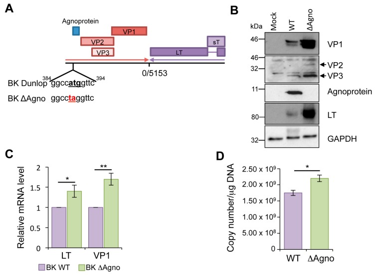
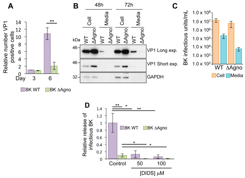
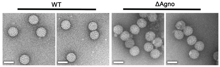
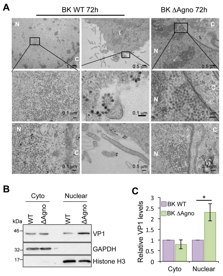
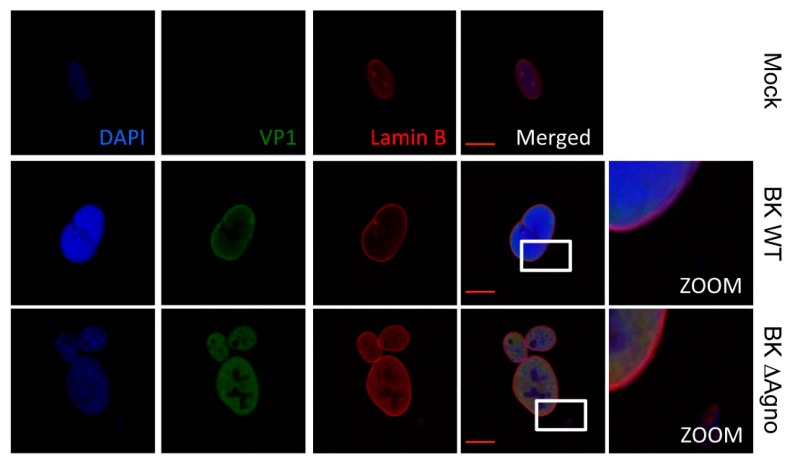
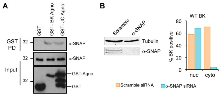
BK polyomavirus (BKPyV; hereafter referred to as BK) causes a lifelong chronic infection and is associated with debilitating disease in kidney transplant recipients. Despite its importance, aspects of the virus life cycle remain poorly understood. In addition to the structural proteins, the late region of the BK genome encodes for an auxiliary protein called agnoprotein. Studies on other polyomavirus agnoproteins have suggested that the protein may contribute to virion infectivity. Here, we demonstrate an essential role for agnoprotein in BK virus release. Viruses lacking agnoprotein fail to release from host cells and do not propagate to wild-type levels. Despite this, agnoprotein is not essential for virion infectivity or morphogenesis. Instead, agnoprotein expression correlates with nuclear egress of BK virions. We demonstrate that the agnoprotein binding partner α-soluble N-ethylmaleimide sensitive fusion (NSF) attachment protein (α-SNAP) is necessary for BK virion release, and siRNA knockdown of α-SNAP prevents nuclear release of wild-type BK virions. These data highlight a novel role for agnoprotein and begin to reveal the mechanism by which polyomaviruses leave an infected cell.

Keywords: agnoprotein; polyomavirus; virus exit.


Conflict of interest statement

The authors declare no conflict of interest.

Figures

Figure 1
Figure 1
Loss of agnoprotein increases BK gene expression. (A) Schematic illustration of the BK Dunlop genome including the agnoprotein sequence mutated to generate the ΔAgno virus. Agnoprotein start codon in bold and base changes underlined in red; (B) Lysates from RPTE cells transfected with BK WT and ΔAgno genomes were probed with antibodies against early (LT) and late (VP1-3 and agnoprotein) proteins. Glyceraldehyde 3-phosphate dehydrogenase (GAPDH) was included as a protein loading control. Loss of agnoprotein correlated with increased expression of other virus protein products; (C) Levels of early (LT) and late (VP1) mRNA transcripts were measured from RPTE cells containing BK WT or ΔAgno genomes. Levels of virus transcript were increased in the absence of agnoprotein; (D) Virus genome replication was measured by qPCR in RPTE cells containing BK WT and ΔAgno virus. Genome replication was increased in the absence of agnoprotein. All experiments are representative of at least three independent experimental repeats. Significance of changes were analyzed by Student’s t-test and indicated by * p <0.05, ** p <0.01.
Figure 2
Figure 2
Agnoprotein facilitates virion release and enhances virus propagation. (A) RPTE cells transfected with BK WT and ΔAgno genomes were incubated over a 6-day time course, and levels of VP1 protein expression determined by indirect immunofluorescence using Incucyte Zoom software (Essen BioScience, Ann Arbor, MI, USA). Levels of VP1 expression are shown relative to the Day 3 BK WT sample. Significance of the changes were analyzed by Student’s t-test and indicated by ** p <0.01; (B) BK virus lacking agnoprotein fails to release virus into the cell culture media. Whole cell lysates and media samples from RPTE cells transfected with BK WT or ΔAgno genomes were analyzed at 48 and 72 h post-transfection for the VP1 capsid protein. GAPDH served as a protein loading control for the whole cell lysates; (C) RPTE cells were infected with BK WT and ΔAgno and cell-associated and media fractions harvested separately. Fluorescence focus assay was then performed to determine the IU/mL−1 of virus in the cells and supernatant; (D) Effect of the anion channel blocker DIDS is independent of agnoprotein. RPTE cells were infected with BK WT or ΔAgno and treated with dimethyl sulphoxide (DMSO) only (control) or 50–100 μM DIDS at 48 h post infection. Media and cell-associated fractions were harvested separately at 72 h post infection. Infectious virus titers were quantified by fluorescence focus assay on naïve RPTE cells and the proportion of total infectivity released into the media for each condition was calculated. Levels of released infectivity are represented as relative to the untreated BK WT samples. The graph corresponds to an average of three experimental repeats. Significance was analyzed by Student’s t-test and is indicated by an asterix * p <0.05, ** p <0.01.
Figure 3
Figure 3
Loss of agnoprotein does not impair BK virion assembly. Negative stain electron micrograph of BK WT and ΔAgno virions following centrifugation through an isopycnic caesium chloride gradient. Scale bars 50 nm.
Figure 4
Figure 4
Agnoprotein facilitates nuclear release of BK virions. (A) Electron microscopy analysis of BK WT and ΔAgno infected RPTE cells (n = 40 cells). Boxed areas in the upper panel are shown at higher magnification in the middle panels. Viral particles of about 40 nm in diameter were found in the nuclei of BK WT and ΔAgno transfected cells. Nuclei (N) and cytoplasm (C) are labeled. Scale bars are shown in the panels; (B) Cell fractionation of RPTE cells transfected with BK WT or ΔAgno genomes. Fractions were probed with for VP1 expression. Antibodies detecting GAPDH and Histone H3 served as markers for the cytoplasm and nuclear fractions; (C) Quantification of the Western blot data was performed using ImageJ software (1.8.0_101, NIH, USA) on the VP1 positive bands and is represented relative to BK WT VP1. The graph corresponds to an average from three independent experimental repeats. Significance was analyzed by Student’s t-test and is indicated by an asterix * p <0.05.
Figure 5
Figure 5
Lamin B localization is not altered by agnoprotein. Immunofluorescence staining of RPTE cells 72 h post transfection with BK WT or ΔAgno genomes. Cells were incubated with antibodies against VP1 and Lamin B and a secondary antibodies. Alexa Fluor 488 chicken anti-mouse and Alexa Fluor 594 chicken anti-rabbit. 4′,6-diamidino-2-phenylindole (DAPI) was used to indicate cell nuclei. Representative images are shown from at least three independent experimental repeats and white frames indicate area shown in the zoomed image. Scale bar 10 μm.
Figure 6
Figure 6
The agnoprotein binding partner α-SNAP is required for BK virion release. (A) Recombinant GST-agnoprotein interacts with α-SNAP. Bacterial expressed GST-agnoproteins from BK and JC virus bound to glutathione-agarose beads were incubated with RPTE cell lysates. GST alone served as a negative control. Bound samples were probed with an anti-α-SNAP antibody; (B) Quantification of transmission electron microscopy data. RPTE cells infected with BK WT were treated with siRNA targeting α-SNAP or a scrambled control and electron microscopy used to quantify the numbers cells demonstrating BK virions in nuclear (nuc) and cytoplasmic (cyto) compartments from 50 cells. Associated Western blots for α-SNAP to confirm effective knockdown. Tubulin serves as a loading control.

References

    1. Buck C.B., van Doorslaer K., Peretti A., Geoghegan E.M., Tisza M.J., An P., Katz J.P., Pipas J.M., McBride A.A., Camus A.C., et al. The Ancient Evolutionary History of Polyomaviruses. PLoS Pathog. 2016;12:e1005574. doi: 10.1371/journal.ppat.1005574. - DOI - PMC - PubMed
    1. Peretti A., FitzGerald P.C., Bliskovsky V., Pastrana D.V., Buck C.B. Genome Sequence of a Fish-Associated Polyomavirus, Black Sea Bass (Centropristis striata) Polyomavirus 1. Genome Announc. 2015;3:e01476-14. doi: 10.1128/genomeA.01476-14. - DOI - PMC - PubMed
    1. Peretti A., FitzGerald P.C., Bliskovsky V., Buck C.B., Pastrana D.V. Hamburger polyomaviruses. J. Gen. Virol. 2015;96:833–839. doi: 10.1099/vir.0.000033. - DOI - PMC - PubMed
    1. Hurdiss D.L., Morgan E.L., Thompson R.F., Prescott E.L., Panou M.M., Macdonald A., Ranson N.A. New Structural Insights into the Genome and Minor Capsid Proteins of BK Polyomavirus using Cryo-Electron Microscopy. Structure. 2016;24:528–536. doi: 10.1016/j.str.2016.02.008. - DOI - PMC - PubMed
    1. Feng H., Shuda M., Chang Y., Moore P.S. Clonal integration of a polyomavirus in human Merkel cell carcinoma. Science. 2008;319:1096–1100. doi: 10.1126/science.1152586. - DOI - PMC - PubMed

MeSH terms

Substances