. 2018 Mar 19;23(3):687.
doi: 10.3390/molecules23030687.

Miro1 Enhances Mitochondria Transfer from Multipotent Mesenchymal Stem Cells (MMSC) to Neural Cells and Improves the Efficacy of Cell Recovery

Affiliations

Miro1 Enhances Mitochondria Transfer from Multipotent Mesenchymal Stem Cells (MMSC) to Neural Cells and Improves the Efficacy of Cell Recovery

Valentina A Babenko et al. Molecules. .

Abstract

A recently discovered key role of reactive oxygen species (ROS) in mitochondrial traffic has opened a wide alley for studying the interactions between cells, including stem cells. Since its discovery in 2006, intercellular mitochondria transport has been intensively studied in different cellular models as a basis for cell therapy, since the potential of replacing malfunctioning organelles appears to be very promising. In this study, we explored the transfer of mitochondria from multipotent mesenchymal stem cells (MMSC) to neural cells and analyzed its efficacy under normal conditions and upon induction of mitochondrial damage. We found that mitochondria were transferred from the MMSC to astrocytes in a more efficient manner when the astrocytes were exposed to ischemic damage associated with elevated ROS levels. Such transport of mitochondria restored the bioenergetics of the recipient cells and stimulated their proliferation. The introduction of MMSC with overexpressed Miro1 in animals that had undergone an experimental stroke led to significantly improved recovery of neurological functions. Our data suggest that mitochondrial impairment in differentiated cells can be compensated by receiving healthy mitochondria from MMSC. We demonstrate a key role of Miro1, which promotes the mitochondrial transfer from MMSC and suggest that the genetic modification of stem cells can improve the therapies for the injured brain.

Keywords: astrocyte; ischemia; mitochondria; stroke; tunneling nanotubes.


Conflict of interest statement

The authors declare no conflict of interest.

Figures

Figure 1
Figure 1
The transfer of mitochondria from MMSC to astrocytes and neural cells following mitochondrial damage. (A) Mitochondrial transfer from MMSCs to astrocytes. DsRed-labeled mitochondria from MMSC were transferred to astrocytes with GFP-labeled mitochondria. The intracellular location of red fluorescence was confirmed using confocal line-scanning microscope analysis of cells along the z-axis (bottom); (BD) Mitochondrial fragmentation in astrocytes stained with TMRE (tetramethylrhodamine, ethyl ester) after 5 h of oxygen-glucose deprivation (OGD). Averagely shorter mitochondrial fragments (D) supports mitochondrial fragmentation; (E) The efficacy of mitochondrial transfer from MMSС to astrocytes is increased after OGD; (F) The transfer of DsRed-labeled mitochondria from ММСК to ρ0 PC12 cells; (G) MMSCs more efficiently transferred mitochondria to ρ0 PC12 cells than to native PC12 cells. Scale bars = 10 µm (A, B), and 20 µm (F). All experiments were performed at least in triplicate; * denotes significant differences between groups (p < 0.05) (One-way ANOVA, followed by Tukey’s post hoc analysis). Values are given as mean ± standard error of the mean (SEM).
Figure 2
Figure 2
Mitochondria transfer from MMSCs to neural cells is supported by tunneling nanotubes (TNT). Formation of TNT between MMSC with DsRed-labelled mitochondria and unlabeled PC12 cells (A) and MMSC with GFP-labelled mitochondria and DsRed-labelled astrocytes (B); MMSC-derived mitochondria are seen in TNT (arrows). More TNTs are observed after OGD or overexpression of Miro1 in MMSC (C). Scale bars = 20 µm (A,B). All experiments were performed at least in triplicate; *,# denotes significant differences with respect to the MMSC group (p < 0.05) or the MMSC + Astrocytes group, (One-way ANOVA, followed by Tukey’s post hoc). Values are given as mean ± standard error of the mean (SEM).
Figure 3
Figure 3
Retardation of glycolysis and higher proliferation in ρ0 PC12 cells associated with the transfer of mitochondria from MMSC. (A) Co-cultivation of MMSC and ρ0 PC12 cells was associated with lower production of lactate, possibly speeding ATP production from oxidative phosphorylation and blocking glycolysis, thus increasing the normalized cell index (B) and reducing the doubling time (C), which demonstrated the activation of proliferation in PC12 cells. All experiments were performed at least in triplicate; * denotes significant differences between groups (p < 0.05) (One-way ANOVA, followed by Tukey’s post hoc analysis). Values are given as mean ± standard error of the mean (SEM).
Figure 4
Figure 4
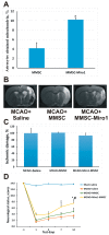
Beneficial effects of Miro1 overexpression on mitochondrial transfer. (A) Efficacy of mitochondrial transfer from MMSC overexpressing Miro1 to astrocytes after 24 h of co-cultivation; (B) Representative T2-weighted MR-images from coronal brain sections obtained 14 days after middle cerebral artery occlusion (MCAO). Hyperintensive regions refer to ischemic areas; (C) The volume of the ischemic lesions in the brain on day 14 after MCAO; (D) Effect of MMSC transplantation on the neurological status at different times after stroke. Intravenous injection with either native MMSCs or MMSC-Miro1 caused a significant decrease of the neurological deficit, with MMSC-Miro1 being more effective. *, p < 0.05 vs. ischemic saline controls; #, p < 0.05 vs. ischemic MMSC-treated rats. All cell culture experiments were performed at least in triplicate, and at least six animals were used for each group in the stroke study.

References

    1. Droge W. Free radicals in the physiological control of cell function. Physiol. Rev. 2002;82:47–95. doi: 10.1152/physrev.00018.2001. - DOI - PubMed
    1. Liao P.C., Tandarich L.C., Hollenbeck P.J. ROS regulation of axonal mitochondrial transport is mediated by Ca2+ and JNK in Drosophila. PLoS ONE. 2017;12:e0178105. doi: 10.1371/journal.pone.0178105. - DOI - PMC - PubMed
    1. Debattisti V., Gerencser A.A., Saotome M., Das S., Hajnoczky G. ROS control mitochondrial motility through p38 and the motor adaptor miro/trak. Cell Rep. 2017;21:1667–1680. doi: 10.1016/j.celrep.2017.10.060. - DOI - PMC - PubMed
    1. Shin B., Cowan D.B., Emani S.M., Del Nido P.J., McCully J.D. Mitochondrial transplantation in myocardial ischemia and reperfusion injury. Adv. Exp. Med. Biol. 2017;982:595–619. - PubMed
    1. Liang X., Ding Y., Zhang Y., Tse H.F., Lian Q. Paracrine mechanisms of mesenchymal stem cell-based therapy: Current status and perspectives. Cell Transplant. 2014;23:1045–1059. doi: 10.3727/096368913X667709. - DOI - PubMed