. 2018 Mar 19;10(3):137.
doi: 10.3390/v10030137.

Beet Necrotic Yellow Vein Virus Noncoding RNA Production Depends on a 5'→3' Xrn Exoribonuclease Activity

Affiliations

Beet Necrotic Yellow Vein Virus Noncoding RNA Production Depends on a 5'→3' Xrn Exoribonuclease Activity

Alyssa Flobinus et al. Viruses. .

Abstract

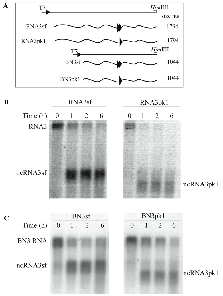
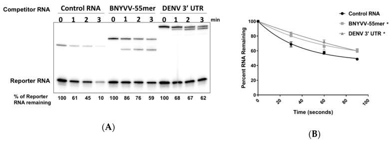
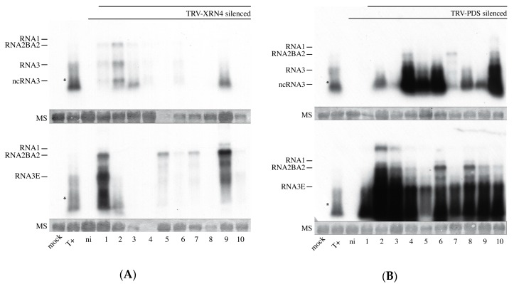
The RNA3 species of the beet necrotic yellow vein virus (BNYVV), a multipartite positive-stranded RNA phytovirus, contains the 'core' nucleotide sequence required for its systemic movement in Beta macrocarpa. Within this 'core' sequence resides a conserved "coremin" motif of 20 nucleotides that is absolutely essential for long-distance movement. RNA3 undergoes processing steps to yield a noncoding RNA3 (ncRNA3) possessing "coremin" at its 5' end, a mandatory element for ncRNA3 accumulation. Expression of wild-type (wt) or mutated RNA3 in Saccharomyces cerevisiae allows for the accumulation of ncRNA3 species. Screening of S.cerevisiae ribonuclease mutants identified the 5'-to-3' exoribonuclease Xrn1 as a key enzyme in RNA3 processing that was recapitulated both in vitro and in insect cell extracts. Xrn1 stalled on ncRNA3-containing RNA substrates in these decay assays in a similar fashion as the flavivirus Xrn1-resistant structure (sfRNA). Substitution of the BNYVV-RNA3 'core' sequence by the sfRNA sequence led to the accumulation of an ncRNA species in yeast in vitro but not in planta and no viral long distance occurred. Interestingly, XRN4 knockdown reduced BNYVV RNA accumulation suggesting a dual role for the ribonuclease in the viral cycle.

Keywords: BNYVV; VIGS; Viral noncoding RNA; exoribonuclease; flavivirus.


Conflict of interest statement

The authors declare no conflict of interest and state that Elodie Klein is an employee of SESV and has provided help with the RNA assays. The founding sponsors had no role in the design of the study; in the collection, analyses, or interpretation of data; in the writing of the manuscript, and in the decision to publish the results.

Figures

Figure 1
Figure 1
Exoribonucleases are responsible for noncoding RNA3 species accumulation in Saccharomyces cerevisiae. (A) Representation of the expression vector cassettes (solid lines) used to produce RNA3 and RNA3E species under the control of constitutive G6PDH promoter (black arrowhead). Capped (•) full-length RNAs and ncRNA species are depicted by waved grey and black lines, respectively. Wild-type (wt) ‘core’ sequence is presented in dark and mutated “core” in grey bold lines. The “coremin” motif and its antisense orientation, “nimeroc”, are depicted by black sense and antisense arrows, respectively. Drawings are not to scale and for a better representation of RNA3 species, refer to figure 2 of reference [27]. RNA sizes are presented on the right; (B,C) Northern blot analyses of RNA3 and ncRNA species produced in yeasts; (B) vectors producing wt RNA3, mutated RNA3E, or control vector (Ø) were introduced in S. cerevisiae (lanes 1–3) or Xrn1 defective strain (Δxrn1) (lanes 4–5). Expression of RNA3 (lanes 6–10) was performed in ∆xrn1 strain together with an empty vector (EV, lane 6) or vectors expressing: yeast Xrn1 (lane 7), plant ATXRN4 (XRN4, lanes 8 and 9), or a defective Xrn1 enzyme (Xrn1cat, lane 10). Total RNAs were subjected to northern blot analysis using a beet necrotic yellow vein virus (BNYVV)-specific 3′ probe complementary to nt 1277–1774; (C) total RNAs from yeast expressing RNA3 were differentially visualized using two probes (lanes 1 and 2). RNA3, RNA3*, ncRNA3, and ncRNA3* were revealed with a coremin-specific probe (lane 1) while RNA3* and ncRNA3* appeared only when the CYC1 terminator-specific probe was used (lane 2). Black triangles indicate the two full-length RNA3 species. Loadings were visualized by ethidium-bromide staining (rRNA).
Figure 2
Figure 2
Accumulation of noncoding RNA3sf from chimeric RNA3sf inhibits Xrn1 and AtXRN4 exoribonucleases in Saccharomyces cerevisiae. (A) The ‘core’ sequence was replaced by a wt (sf) or mutated (pk1) flavivirus sequence to produce RNA3sf and RNA3pk1, respectively. PK1 pseudoknot involving stem-loop II (SL-II) is shown; (B) northern blot analyses of RNA3 and ncRNA species produced in yeasts. Plasmids allowing the expression of RNA species indicated that RNA3, RNA3E, RNA3sf, RNA3pk1, or empty vector (Ø) were introduced in wt yeasts (lanes 1–5) or Xrn1-defective yeasts (Δxrn1, lanes 6–9) complemented with a vector allowing for the production of yeast Xrn1 (lanes 6, 8, and 9) or plant XRN4 (lane 7). The RNA3* and ncRNA3* are similar to RNA3 and ncRNA3, respectively, but possess a CYC1 terminator sequence followed by a polyA tail (see Figure 1 and text for details). Positions of the RNA species are indicated on the right. Total RNAs were subjected to northern blot analysis using a BNYVV-specific 3′ probe complementary to nt 1277–1774. The partial complementarity of the probe with the ncRNA3sf species does not allow for quantitative comparisons. Loading was visualized by ethidium-bromide staining (rRNA). No sample was loaded between lanes 8 and 9.
Figure 3
Figure 3
Both ncRNA3sf and ncRNA3pk1 stall Xrn1 processing in vitro. (A) Schematic representation of the T7-driven cDNA clones constructs used to produce run-off in vitro transcripts depicted in waved lines. Double and single arrowheads correspond to the sf and pk1 structural motifs, respectively. The position of the restriction sites used and the size of the transcripts are indicated; (B) 5′ phosphorylated chimeric RNA3sf or RNA3pk1 and (C) BN3sf or BN3pk1 species were mixed with commercial Xrn1 enzyme for 6 h. Aliquots were sampled at the time indicated and RNAs species were detected by northern blot using a specific DNA probe able to reveal both full-length and ncRNA3 species.
Figure 4
Figure 4
Characterization of the minimal RNA3 3′ domain required for the efficient stalling of the Xrn1 enzyme. (A) Representation of the fragments obtained using T7 promoter containing primer (T7-RNA3F) and reverse primers (positions specified by blue boxes) cloned into pUC57 that served as templates for the production of 1R to 9R transcripts, ranging from 989 to 558 nt, possessing the same 5′ extremity with decreasing 3′ end length. The arrowhead locates the 5′ position of the ncRNA3 species (nt 1234). The expected product size after RppH and Xrn1 treatments are specified. Construct 6R was not obtained; (B) after 6 h of RppH/Xrn1 treatment, RNAs were analyzed by a 24-cm long run on 1.5% denaturing-agarose gel followed by northern blot using a radiolabeled DNA oligomer probe complementary to the “coremin” sequence. The ncRNA3 transcripts and 1R transcripts were treated with RppH alone or with RppH and Xrn1. The lengths of some RNA species are specified on the left side.
Figure 5
Figure 5
A 55 nt fragment containing the coremin motif is sufficient to stall XRN1. (A) 5′ monophosphorylated radiolabeled RNA substrates containing either control sequences derived from pGEM-4 or a 101-nt RNA containing a 55 base fragment from the RNA3 segment of BNYVV (nts 1222–1277) at the 3′ end and 53 nts of pGEM-4 polylinker sequence at its 5′ end to serve as a landing site for 5′-to-3′ exonucleases) were incubated with either purified recombinant XRN1 from Kluyveromyces lactis (rXrn1 panel) or cytoplasmic extract from C6/36 Aedes albopictus cells for the times indicated. Reaction products were resolved on a 5% acrylamide gel containing urea and viewed by phosphorimaging. (B) Top: sequence of the 55 nts BNYVV RNA fragment. The black arrow indicates the site of XRN1 stalling. Fragments B-1, B-2, and B-3 containing BNYVV-specific sequences ranging from position 1222 to the base indicated in the figure. Bottom: 5′ monophosphorylated radiolabeled RNA substrates containing either control sequences derived from pGEM-4 or the B-1, B-2, or B-3 fragments of the RNA3 segment (nts 1222–1277) as indicated in the top part of the panel (inserted into the pGEM-4 polylinker as indicated in panel A) were incubated with purified recombinant XRN1 for the times indicated. Reaction products were resolved on a 5% acrylamide gel containing urea and viewed by phosphorimaging. Arrows indicate the positions of the RNA species stalling XRN1.
Figure 6
Figure 6
A 55 nt core fragment of RNA3 of BNYVV represses Xrn1. (A) A radiolabeled RNA containing a 5′ monophosphate (Reporter) was incubated with purified recombinant XRN1 for the times indicated. A 20X molar excess of lightly radiolabeled, 5′ monophosphorylated non-specific competitor RNA (‘Control RNA’ lanes), a competitor transcript containing the 55 nt core BNYVV RNA3 fragment (‘BNYVV-55mer’ lanes), or a competitor transcript containing the 3′ UTR of Dengue virus type 2 (‘DENV 3′ UTR’ lanes) was added to reactions. After the times indicated, reaction products were analyzed on 5% polyacrylamide gels containing urea and visualized by phosphorimaging. (B) Graphical presentation of the effect of the various competitor RNAs on Xrn1 activity on the Reporter transcript. Results shown are from three independent experiments. The asterisk represents a p value of <0.001 at both time points for viral 3′ UTR/BNYVV-55mer compared to the control as determined using a Turkey’s multiple comparisons test as a post-hoc test.
Figure 7
Figure 7
The BNYVV replication machinery does not replicate RNA3sf and poorly replicates RNA3pk1. Chenopodium quinoa leaves were rub-inoculated (A) or protoplasts electroporated (B) with RNA1+2 alone (-) or supplemented with RNA3, RNA3sf, or RNA3pk1. (A) RNA contents of local lesions were analyzed 7 days post-inoculation (dpi) by northern blot using BNYVV-RNA-specific radiolabeled probes. RNA3pk1 loading was three times lower than the other samples; the membrane was exposed accordingly and visualized with a split line. (B) Protoplasts contents were analyzed 40 h post-inoculation (hpi) as for (A). Loadings are visualized by ethidium-bromide staining of total RNAs (rRNA). NI, non-infected.
Figure 8
Figure 8
TRV-mediated VIGS of XRN4 is efficient in leaves. RT qPCR analyses of XRN4 mRNA within leaves from non-infected plants (WT NI), TRV-XRN (XRN), and TRV-PDS (PDS) silenced N. benthamiana leaves (NI) or rub-inoculated with RNA1+2BA2 supplemented with RNA3 (BA2+3) or RNA3E (BA2+3E). Non-silenced N. benthamiana inoculated with RNA1+2BA2 supplemented with RNA3 served as control (WT BA2+3). Values were normalized to a mock treated control plant (WT NI). Quantitative PCR was performed using specific primers targeting the 5′ (blue) and the 3′ (red) region flanking the XRN4 trigger sequence present in TRV RNA2 and GAPDH and PP2A cellular RNAs. The numbers of sampled plant are specified (N). Student test p-values were >0.005 (a) or <0.005 (b) using WT NI as reference (upper letter) or comparing 5′ and 3′ relative expression (lower letter).
Figure 9
Figure 9
BNYVV RNA accumulation is compromised in XRN4 KD plants. TRV-XRN- (A) and TRV-PDS- (B) silenced N. benthamiana plants were rub-inoculated with RNA1+2BA2 supplemented with RNA3 (top) or RNA3E (bottom). Equivalent amount of total RNAs (~10 µg, visualized by methylene blue staining, MS) were extracted from the inoculated leaf of each plant (1–10) and analysed by northern blot using specific probes for BNYVV RNA1, 2, and 3. Mock, control plant; T+, N. benthamiana infected with RNA1+2BA2+RNA3; ni, TRV-silenced and non-inoculated with BNYVV. The poor quality of the northern blots is due to mixed TRV and BNYVV infection. Samples in panel A lanes 5 (top) and 4 (bottom) were lost. The asterisk indicates the position of ncRNA3. Upper bands of panel B (e.g., lanes 2 and 3) are of unknown origin and may correspond to uncompleted denaturation of viral RNAs. Panel A and B membrane treatments were simultaneous using the same combination and amount of probes. Both blots were exposed 120 h to allow the visualization of viral RNAs of panel A.

Similar articles

Cited by

References

    1. Schoenberg D.R., Maquat L.E. Regulation of cytoplasmic mRNA decay. Nat. Rev. Genet. 2012;13:246–259. doi: 10.1038/nrg3160. - DOI - PMC - PubMed
    1. Łabno A., Tomecki R., Dziembowski A. Cytoplasmic RNA decay pathways—Enzymes and mechanisms. Biochim. Biophys. Acta. 2016;1863:3125–3147. doi: 10.1016/j.bbamcr.2016.09.023. - DOI - PubMed
    1. Rozovics J.M., Chase A.J., Cathcart A.L., Chou W., Gershon P.D., Palusa S., Wilusz J., Semler B.L. Picornavirus modification of a host mRNA decay protein. mBio. 2012;3:e00431-12. doi: 10.1128/mBio.00431-12. - DOI - PMC - PubMed
    1. Sokoloski K.J., Dickson A.M., Chaskey E.L., Garneau N.L., Wilusz C.J., Wilusz J. Sindbis virus usurps the cellular hur protein to stabilize its transcripts and promote productive infections in mammalian and mosquito cells. Cell Host Microbe. 2010;8:196–207. doi: 10.1016/j.chom.2010.07.003. - DOI - PMC - PubMed
    1. Csorba T., Kontra L., Burgyán J. Viral silencing suppressors: Tools forged to fine-tune host-pathogen coexistence. Virology. 2015;479–480:85–103. doi: 10.1016/j.virol.2015.02.028. - DOI - PubMed

Publication types

Supplementary concepts

LinkOut - more resources