Extracellular Vesicles Derived from Wharton's Jelly Mesenchymal Stem Cells Prevent and Resolve Programmed Cell Death Mediated by Perinatal Hypoxia-Ischemia in Neuronal Cells
- PMID: 29562785
- PMCID: PMC6434490
- DOI: 10.1177/0963689717738256
Extracellular Vesicles Derived from Wharton's Jelly Mesenchymal Stem Cells Prevent and Resolve Programmed Cell Death Mediated by Perinatal Hypoxia-Ischemia in Neuronal Cells
Abstract
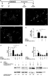
Hypoxic-ischemic (HI) insult in the perinatal phase harbors a high risk of encephalopathy in the neonate. Brain cells undergo apoptosis, initiating neurodegeneration. So far, therapeutic approaches such as cooling remain limited. Transplantation of mesenchymal stem cells (MSCs) exhibits therapeutic success despite the short-time survival in the host brain, providing strong evidence that their beneficial effects are largely based on secreted factors, including extracellular vesicles (EVs). The aim of this study was to investigate the effects of human Wharton's jelly MSC (hWJ-MSC)-derived EVs on neuroprotection and neuroregeneration, using an in vitro model of oxygen-glucose deprivation/reoxygenation (OGD/R) mimicking HI injury in the mouse neuroblastoma cell line neuro2a (N2a). hWJ-MSC-derived EVs were isolated from cell culture supernatants by multistep centrifugation and identified by endosomal marker expression and electron microscopy. OGD/R significantly increased DNA fragmentation and caspase 3 ( Casp3) transcription in N2a cells relative to undamaged cells. OGD/R-mediated DNA fragmentation and Casp3 expression could be prevented as well as resolved by the addition of hWJ-MSC-derived EV before and after OGD, respectively. hWJ-MSC-derived EV also tended to increase the phosphorylation of the B cell lymphoma 2 (Bcl2) family member Bcl-2-antagonist of cell death (BAD) in N2a cells, when added prior or post OGD, thereby inactivating the proapoptotic function of BAD. Fluorescence confocal microscopy revealed the close localization of hWJ-MSC-derived EVs to the nuclei of N2a cells. Furthermore, EVs released their RNA content into the cells. The expression levels of the microRNAs (miRs) let-7a and let-7e, known regulators of Casp3, were inversely correlated to Casp3. Our data suggest that hWJ-MSC-derived EVs have the potential to prevent and resolve HI-induced apoptosis in neuronal cells in the immature neonatal brain. Their antiapoptotic effect seems to be mediated by the transfer of EV-derived let-7-5p miR.
Keywords: apoptosis; extracellular vesicles (EVs); let-7-5p; neuroprotection; neuroregeneration; oxygen–glucose deprivation/reoxygenation (OGD/R).
Conflict of interest statement
Figures


References
-
- Johnston MV, Trescher WH, Ishida A, Nakajima W. Neurobiology of hypoxic-ischemic injury in the developing brain. Pediatric Res. 2001;49(6):735–741. - PubMed
-
- Vannucci SJ, Hagberg H. Hypoxia-ischemia in the immature brain. J Exp Biol. 2004;207(Pt 18):3149–3154. - PubMed
-
- Zhu C, Wang X, Xu F, Bahr BA, Shibata M, Uchiyama Y, Hagberg H, Blomgren K. The influence of age on apoptotic and other mechanisms of cell death after cerebral hypoxia-ischemia. Cell Death Differ. 2005;12(2):162–176. - PubMed
Publication types
MeSH terms
Substances
LinkOut - more resources
Full Text Sources
Other Literature Sources
Research Materials

