

Characterization of association of human mitochondrial lysyl-tRNA synthetase with HIV-1 Pol and tRNA3Lys
- PMID: 29562886
- PMCID: PMC5863373
- DOI: 10.1186/s12858-018-0092-x
Characterization of association of human mitochondrial lysyl-tRNA synthetase with HIV-1 Pol and tRNA3Lys
Abstract
Background: An important step in human immunodeficiency virus type 1 (HIV-1) replication is the packaging of tRNA3Lys from the host cell, which plays the role of primer RNA in the process of initiation of reverse transcription. The viral GagPol polyprotein precursor, and the human mitochondrial lysyl-tRNA synthetase (mLysRS) from the host cell, have been proposed to be involved in the packaging process. More specifically, the catalytic domain of mLysRS is supposed to interact with the transframe (TF or p6*) and integrase (IN) domains of the Pol region of the GagPol polyprotein.
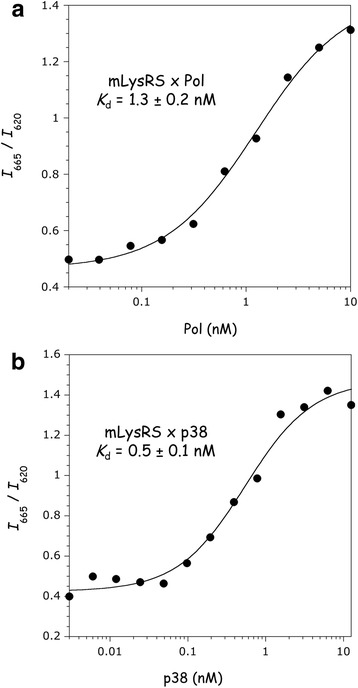
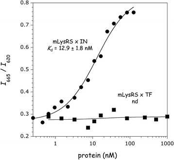
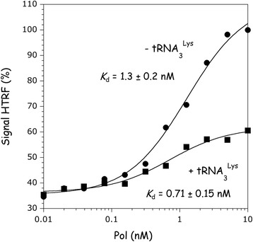
Results: In this work, we report a quantitative characterization of the protein:protein interactions between mLysRS and its viral partners, the Pol polyprotein, and the isolated integrase and transframe domains of Pol. A dissociation constant of 1.3 ± 0.2 nM was determined for the Pol:mLysRS interaction, which exemplifies the robustness of this association. The protease and reverse transcriptase domains of GagPol are dispensable in this association, but the TF and IN domains have to be connected by a linker polypeptide to recapitulate a high affinity partner for mLysRS. The binding of the viral proteins to mLysRS does not dramatically enhance the binding affinity of mLysRS for tRNA3Lys.
Conclusions: These data support the conclusion that the complex formed between GagPol, mLysRS and tRNA3Lys, which involves direct interactions between the IN and TF domains of Pol with mLysRS, is more robust than suggested by the previous models supposed to be involved in the packaging of tRNA3Lys into HIV-1 particles.
Keywords: Binding affinity; HIV-1; Integrase; Mitochondrial lysyl-tRNA synthetase; Transframe (TF or p6*); tRNA3 Lys packaging.
Conflict of interest statement
Ethics approval and consent to participate
Not applicable.
Consent for publication
Not applicable.
Competing interests
The authors declare that they have no competing interests.
Publisher’s Note
Springer Nature remains neutral with regard to jurisdictional claims in published maps and institutional affiliations.
Figures


Similar articles
-
How HIV-1 Integrase Associates with Human Mitochondrial Lysyl-tRNA Synthetase.Viruses. 2020 Oct 21;12(10):1202. doi: 10.3390/v12101202. Viruses. 2020. PMID: 33096929 Free PMC article.
-
Association of mitochondrial Lysyl-tRNA synthetase with HIV-1 GagPol involves catalytic domain of the synthetase and transframe and integrase domains of Pol.J Mol Biol. 2011 Jul 29;410(5):875-86. doi: 10.1016/j.jmb.2011.03.005. J Mol Biol. 2011. PMID: 21763493
-
Association of human mitochondrial lysyl-tRNA synthetase with HIV-1 GagPol does not require other viral proteins.Biochim Open. 2016 Mar 5;2:52-61. doi: 10.1016/j.biopen.2016.02.004. eCollection 2016 Jun. Biochim Open. 2016. PMID: 29632838 Free PMC article.
-
Formation of the tRNALys packaging complex in HIV-1.FEBS Lett. 2010 Jan 21;584(2):359-65. doi: 10.1016/j.febslet.2009.11.038. FEBS Lett. 2010. PMID: 19914238 Free PMC article. Review.
-
The selective packaging and annealing of primer tRNALys3 in HIV-1.Curr HIV Res. 2004 Apr;2(2):163-75. doi: 10.2174/1570162043484988. Curr HIV Res. 2004. PMID: 15078180 Review.
Cited by
-
Aminoacyl-tRNA Synthetase: A Non-Negligible Molecule in RNA Viral Infection.Viruses. 2022 Mar 15;14(3):613. doi: 10.3390/v14030613. Viruses. 2022. PMID: 35337020 Free PMC article. Review.
-
Effect of Lysyl-tRNA Synthetase on the Maturation of HIV-1 Reverse Transcriptase.ACS Omega. 2020 Jun 30;5(27):16619-16627. doi: 10.1021/acsomega.0c01449. eCollection 2020 Jul 14. ACS Omega. 2020. PMID: 32685828 Free PMC article.
-
Hijacking tRNAs From Translation: Regulatory Functions of tRNAs in Mammalian Cell Physiology.Front Mol Biosci. 2020 Dec 17;7:610617. doi: 10.3389/fmolb.2020.610617. eCollection 2020. Front Mol Biosci. 2020. PMID: 33392265 Free PMC article. Review.
-
Interplay between protease and reverse transcriptase dimerization in a model HIV-1 polyprotein.Protein Sci. 2024 Jul;33(7):e5080. doi: 10.1002/pro.5080. Protein Sci. 2024. PMID: 38896002 Free PMC article.
-
How HIV-1 Integrase Associates with Human Mitochondrial Lysyl-tRNA Synthetase.Viruses. 2020 Oct 21;12(10):1202. doi: 10.3390/v12101202. Viruses. 2020. PMID: 33096929 Free PMC article.
References
-
- Gabor J, Cen S, Javanbakht H, Niu MJ, Kleiman L. Effect of altering the tRNA3Lys concentration in human immunodeficiency virus type 1 upon its annealing to viral RNA, GagPol incorporation, and viral infectivity. J Virol. 2002;76(18):9096–9102. doi: 10.1128/JVI.76.18.9096-9102.2002. - DOI - PMC - PubMed
Publication types
MeSH terms
Substances
LinkOut - more resources
Full Text Sources
Other Literature Sources
Research Materials
Miscellaneous
