

Left ventricular hypertrophy diagnosed after a stroke: a case report
- PMID: 29562910
- PMCID: PMC5863476
- DOI: 10.1186/s13256-018-1592-4
Left ventricular hypertrophy diagnosed after a stroke: a case report
Abstract
Background: Stroke is a recognized clinical course of hypertrophic cardiomyopathy. This interesting case showed notable difference on the electrocardiogram of a patient 4 months prior to suffering a stroke and 10 days after suffering a stroke. The pre-stroke electrocardiogram showed atrial fibrillation with a narrow QRS complex, while the post-stroke electrocardiogram showed marked left ventricular hypertrophy. Left ventricular hypertrophy was diagnosed using the Sokolow-Lyon indices. The development of left ventricular hypertrophy a few days after suffering a stroke has not previously been reported.
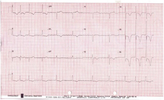
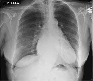
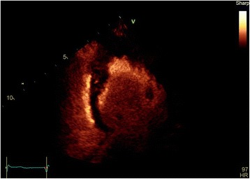
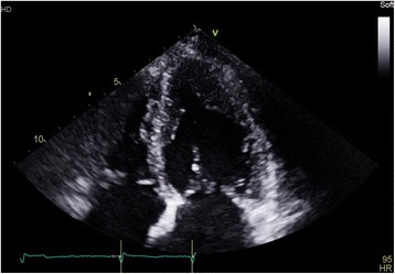
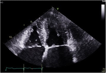
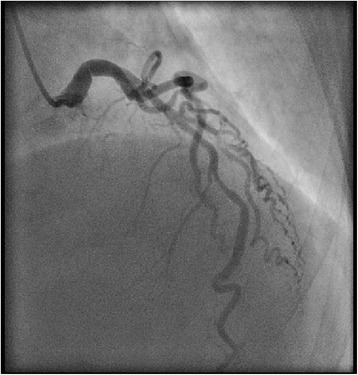
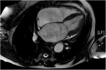
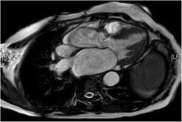
Case presentation: An 83-year-old white British woman with a background history of permanent atrial fibrillation, hypertension, and previous stroke attended the emergency department with a 2-day history of exertional dyspnea, and chest tightness. On examination, she had bibasal crepitations with a systolic murmur loudest at the apex. In-patient investigations include an electrocardiogram, blood tests, chest X-ray, contrast echocardiogram, coronary angiogram, and cardiovascular magnetic resonance imaging. An electrocardiogram showed atrial fibrillation, with inferolateral T wave inversion, and left ventricular hypertrophy. A chest X-ray showed features consistent with pulmonary edema. A contrast echocardiogram showed marked hypertrophy of the mid to apical left ventricle, appearance consistent with apical hypertrophic cardiomyopathy. Coronary angiography showed eccentric shelf-type plaque with non-flow-limiting stenosis in the left coronary artery main stem. Cardiovascular magnetic resonance imaging reported findings highly suggestive of apical hypertrophic cardiomyopathy. Our patient was treated and discharged on rivaroxaban, bisoprolol, and atorvastatin with a follow-up in the cardiomyopathy outpatient clinic.
Conclusions: Electrocardiogram diagnosis of left ventricular hypertrophy led to the diagnosis of apical hypertrophic cardiomyopathy in this patient. Left ventricular hypertrophy was only evident a few days after our patient suffered a stroke. The underlying mechanisms responsible for this remain unclear. Furthermore, differential diagnosis of hypertrophic cardiomyopathy should be considered in people with electrocardiogram criteria for left ventricular hypertrophy. Cardiovascular magnetic resonance imaging is an important diagnostic tool in identifying causes of left ventricular hypertrophy. Family screening should be recommended in patients with new diagnosis of hypertrophic cardiomyopathy.
Keywords: Electrocardiogram; Left ventricular hypertrophy; Stroke.
Conflict of interest statement
Ethics approval and consent to participate
Not applicable.
Consent for publication
Written informed consent was obtained from the patient for publication of this case report and any accompanying images. A copy of the written consent is available for review by the Editor-in-Chief of this journal.
Competing interests
The authors declare that they have no competing interests.
Publisher’s Note
Springer Nature remains neutral with regard to jurisdictional claims in published maps and institutional affiliations.
Figures

Similar articles
-
Call a Spade a Spade: Missed Diagnosis of Apical Hypertrophic Cardiomyopathy.Am J Med Sci. 2019 Oct;358(4):299-303. doi: 10.1016/j.amjms.2019.07.002. Epub 2019 Jul 13. Am J Med Sci. 2019. PMID: 31353027
-
Apical hypertrophic cardiomyopathy with hemodynamically unstable ventricular arrhythmia - Atypical presentation.Indian Heart J. 2016 Sep;68 Suppl 2(Suppl 2):S202-S206. doi: 10.1016/j.ihj.2015.08.011. Epub 2016 Jan 14. Indian Heart J. 2016. PMID: 27751289 Free PMC article.
-
Left ventricular apical hypertrophy in a transplanted heart: a case report.BMC Cardiovasc Disord. 2019 Apr 3;19(1):81. doi: 10.1186/s12872-019-1069-4. BMC Cardiovasc Disord. 2019. PMID: 30943916 Free PMC article.
-
A Cautionary Tale of Hypertrophic Cardiomyopathy-From "Benign" Left Ventricular Hypertrophy to Stroke, Atrial Fibrillation, and Molecular Genetic Diagnostics: A Case Report and Review of Literature.Int J Mol Sci. 2024 Aug 29;25(17):9385. doi: 10.3390/ijms25179385. Int J Mol Sci. 2024. PMID: 39273332 Free PMC article. Review.
-
Apical hypertrophic cardiomyopathy--case report and review of the literature.Georgian Med News. 2013 Mar;(216):19-23. Georgian Med News. 2013. PMID: 23567303 Review.
References
-
- Maron SM, McKenna JW, Downey CB. Hypertrophic cardiomyopathy: clinical manifestations, diagnosis, and evaluation. Uptodate 2017.
-
- Goldberger AL, Mirvis DM, Saperia GM. Left ventricular hypertrophy. Uptodate 2016.
Publication types
MeSH terms
Substances
LinkOut - more resources
Full Text Sources
Other Literature Sources
Medical
Miscellaneous
