. 2018 May 25;122(11):1517-1531.
doi: 10.1161/CIRCRESAHA.118.312789. Epub 2018 Mar 21.

Protein S-Nitrosylation Controls Glycogen Synthase Kinase 3β Function Independent of Its Phosphorylation State

Affiliations

Protein S-Nitrosylation Controls Glycogen Synthase Kinase 3β Function Independent of Its Phosphorylation State

Sheng-Bing Wang et al. Circ Res. .

Abstract

Rationale: GSK-3β (glycogen synthase kinase 3β) is a multifunctional and constitutively active kinase known to regulate a myriad of cellular processes. The primary mechanism to regulate its function is through phosphorylation-dependent inhibition at serine-9 residue. Emerging evidence indicates that there may be alternative mechanisms that control GSK-3β for certain functions.

Objectives: Here, we sought to understand the role of protein S-nitrosylation (SNO) on the function of GSK-3β. SNO-dependent modulation of the localization of GSK-3β and its ability to phosphorylate downstream targets was investigated in vitro, and the network of proteins differentially impacted by phospho- or SNO-dependent GSK-3β regulation and in vivo SNO modification of key signaling kinases during the development of heart failure was also studied.

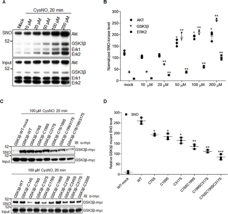
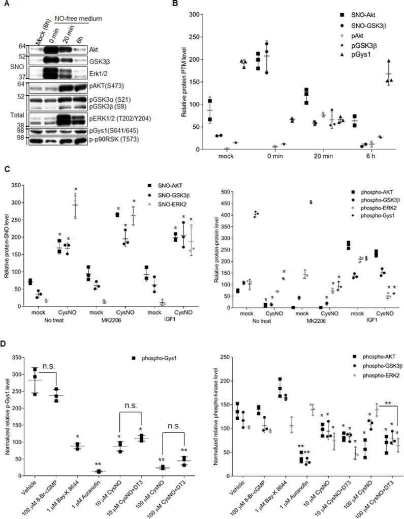
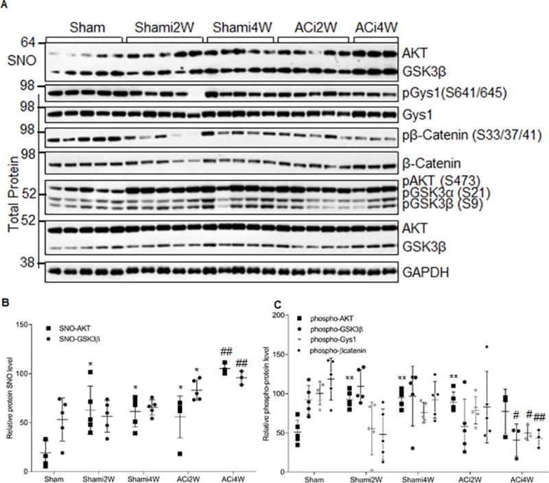
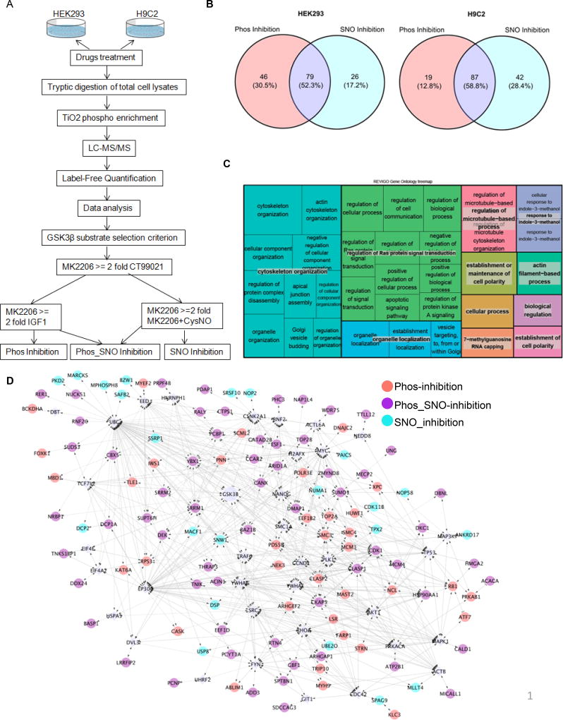
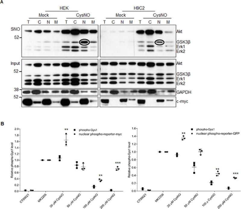
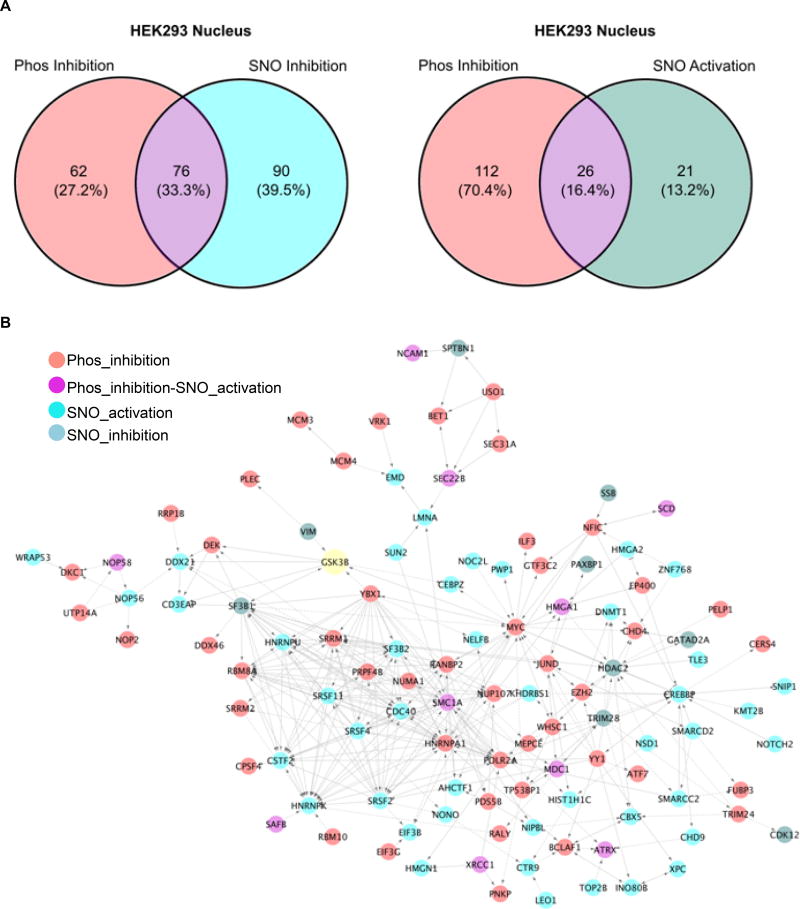
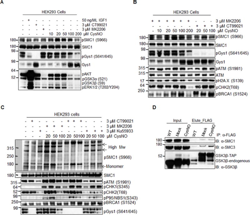
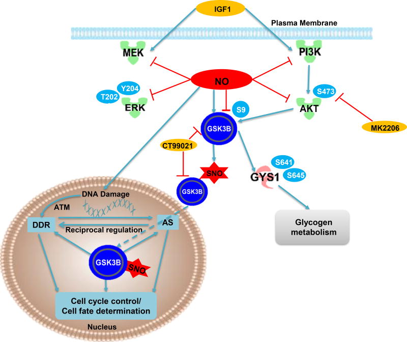
Methods and results: We found that GSK-3β undergoes site-specific SNO both in vitro, in HEK293 cells, H9C2 myoblasts, and primary neonatal rat ventricular myocytes, as well as in vivo, in hearts from an animal model of heart failure and sudden cardiac death. S-nitrosylation of GSK-3β significantly inhibits its kinase activity independent of the canonical phospho-inhibition pathway. S-nitrosylation of GSK-3β promotes its nuclear translocation and access to novel downstream phosphosubstrates which are enriched for a novel amino acid consensus sequence motif. Quantitative phosphoproteomics pathway analysis reveals that nuclear GSK-3β plays a central role in cell cycle control, RNA splicing, and DNA damage response.

Conclusions: The results indicate that SNO has a differential effect on the location and activity of GSK-3β in the cytoplasm versus the nucleus. SNO modification of GSK-3β occurs in vivo and could contribute to the pathobiology of heart failure and sudden cardiac death.

Keywords: S-nitrosylation; glycogen synthase kinase 3 beta; kinase-substrates interactome; nuclear translocation; redox regulation.


Figures

Figure 1
Figure 1. GSK3β is S-nitrosylated at multiple Cys residues in vitro with NO donor treatment
A) Dose dependent induction of S-nitrosylation of GSK3β in vitro with CysNO treatment in HEK293 cells. B) Quantification of A). SNO level is normalized to the input used for biotin switch assay. Error bars represent s.d. of 3 independent experiments. *p<0.02 and **p<0.005 versus mock treatment, using unpaired two-tailed Student’s t test. C) Site-specific S-nitrosylation of GSK3β in HEK 293 cells. Western blots show extent of SNO modification of GSK3β for WT and various single, double or triple GSK3β Cys mutants after CysNO treatment (100 µM for 20 min at 37°C). D) Quantification of the SNO level of GSK3β WT and Cys mutants as shown in the upper panel of (B). SNO levels of each kinase were normalized to their inputs for the biotin switch assay. Error bars represent s.d. of 3 independent experiments. *p<0.05, **p<0.01 and ***p<0.001 versus WT; ##p<0.002 versus the triple mutant C76S/C199S/C317S, using unpaired two-tailed Student’s t test.
Figure 2
Figure 2. SNO reversibly modulates GSK3β kinase activity independent of pSer9 status
A) SNO of kinases (upper) and phosphorylation status of the kinases and their substrates in response to CysNO treatment. HEK293 cells were serum starved for 18 h and treated with 100 µM CysNO for 20 min. CysNO was then removed (0 min) and fresh medium with serum was added and incubated for 20 min or 6 h. B) Quantification of S-nitrosylated kinases, phosphorylation of kinases and their substrates from data obtained as in (A). C) Quantitative analysis of SNO modification of the kinases and the phosphorylation level of kinases/substrates from data obtained as shown in Figure S2B. S-nitrosylation of kinases was normalized to their inputs for the biotin switch assay. Phosphorylation of kinases/substrates was normalized to their total protein levels in the sample. HEK293 cells were serum starved for 18h and pretreated with 3 µM MK2206 or 50 ng/mL IGF1 for 1h to inhibit or activate Akt signaling, respectively. Then CysNO was added to a final concentration at 100 µM and incubated for another 1h. Error bars represent s.d. of 3 independent experiments. *p<0.005 versus mock (CysNO absent) in the same pretreatment group, using unpaired two-tailed Student’s t test.D) SNO inhibition of GSK3β kinase activity is independent of NO-sGC-cGMP-PKG pathway in NRVMs. Quantitative analysis of the phosphorylation level of kinases (right panel) and substrate (left panel) from data obtained as shown in online Figure IIC. NRVM were serum starved for 24 h and treated with vehicle or 1 µM Bay-K 8644, 1 µM auranofin for 1h and 100 µM 8-Br-cGMP for 15 min. For CysNO treatment, the cells were pretreated with or without 1 µM DT3 for 1h. Then CysNO was added to a final concentration at 10 or 100 µM and incubated for another 1h. Phospho-protein level is normalized to its total protein level as relative phospho-protein level. Error bars represent s.d. of 3 independent experiments. *p<0.05 and **p<0.005 versus vehicle treatment group, n.s., not significant, using unpaired two-tailed Student’s t test.
Figure 3
Figure 3. GSK3β is S-nitrosylated in vivo in a guinea pig HF model
A) S-nitrosylation of GSK3β, phosphorylation status of the signaling kinases, and phosphorylation of their substrates at different stages of HF (ACi2W: compensated hypertrophy phase 2 weeks post aortic banding; ACi4W: decompensated heart failure phase 4 weeks post banding) and their non-failing controls (Sham: sham operation with no daily isoproterenol; Shami: sham operation with daily isoproterenol injection). Total heart lysates were used for detection of S-nitrosylation of kinases by biotin switch assay. The phosphorylation status of the kinases and their substrates were detected with phospho-specific antibodies. B) Quantitative analysis of the SNO levels of kinases from data obtained as in (A). SNO kinases were normalized to their inputs for biotin switch assay. Error bars represent s.d. of n=3–5 animals as shown in (A). *p<0.02 versus the Sham control group and ##p<0.003 versus the Shami4W control group using two-tailed unpaired Student’s t test. C, Quantitative analysis of the phosphorylation level of the kinases and known GSK3β substrates from data obtained as in (A). Phosphorylation of kinases/substrates is normalized to their total protein levels in the sample. Error bars represent s.d. of n=3–5 animals as shown in (A). **p<0.002 versus the Sham control group and #p<0.05, ##p<0.003 versus the Shami4W control group using two-tailed unpaired Student’s t test.
Figure 4
Figure 4. SNO modulates the GSK3β kinase–substrate network
A) Work flow showing the design of the quantitative phospho-proteomic approach to define the GSK3β kinase–substrate network. B) Venn diagram showing the distribution of GSK3β substrates modulated by phospho- or SNO-dependent regulation in HEK293 and H9C2 cells. C) REViGO Tree Map showing biological processes enriched for all GSK3β substrates detected in H9C2 cells. Size of the rectangles reflect the p-value of the GO term in the underlying GO annotation database. D) Interactome analysis of GSK3β substrates from HEK293 cells. All candidates, together with GSK3β itself, were up-loaded simultaneously into Cytoscape Reactome FI, and linker genes were allowed. Diamonds, linker genes; circles, observed candidates.
Figure 5
Figure 5. SNO induces GSK3β nuclear translocation
A) Subcellular fractionation blot showing that SNO induces GSK3β translocation to the nuclear fraction and that nuclear GSK3β is S-nitrosylated. Circled bands corresponding to S-nitrosylated GSK3β. B) Quantification of the phosphorylation of the GSK3β nuclear activity reporter and the native GSK3β substrate pGys1 as represented in Figure S4C. Phosphorylation level was normalized to its corresponding total protein level in the sample. Error bars represent s.d. of 3 independent experiments. **p<0.01 and ***p<0.001 versus pGys1 with the same dose of NO treatment, using unpaired two-tailed Student’s t test.
Figure 6
Figure 6. GSK3β nuclear kinase–substrate network
A) Venn diagram showing the distribution of nuclear GSK3β substrates from HEK293 cells. B) Interactome analysis of nuclear GSK3β phospho-substrates from HEK293 cells. All candidates, together with GSK3β itself, were up-loaded simultaneously into Cytoscape Reactome FI, only clustered candidates are shown.
Figure 7
Figure 7. Validation of SMC1A as a GSK3β nuclear substrate
A) Concentration - dependent phosphorylation of SMC1A at Ser966 upon CysNO treatment in HEK293 cells. B) Western blot showing that the CysNO-induced phosphorylation of Ser966 of SMC1A is GSK3β dependent. C) Kinase inhibitor assay showing that both ATM and GSK3β kinases can phosphorylate Ser966 of SMC1A, while induction of phosphorylated high molecular weight product of SMC1A is specific to GSK3β. D) Co-immuno-purfication experiment showing that SMC1A is physically associated with GSK3β in HEK293 cells. TAP-tagged native GSK3β was created in HEK293 cells using CRISPR/Cas9 as described in Online methods. Total cell lysates were used for Co-immuno-purfication.
Figure 8
Figure 8. A working model of the pivotal role of NO on GSK3β function
Based on our data in this study, we hypothesis that NO can induce S-nitrosylation of GSK3β which significantly inhibit its kinase activity while S-nitrosylated GSK3β promote its nuclear translocation where it can interact with splicing machinery to coordinate DDR and cell cycle regulation.

Comment in

Similar articles

Cited by

References

    1. Juhaszova M, Zorov DB, Yaniv Y, Nuss HB, Wang S, Sollott SJ. Role of glycogen synthase kinase-3beta in cardioprotection. Circ Res. 2009;104:1240–1252. - PMC - PubMed
    1. Beurel E, Grieco SF, Jope RS. Glycogen synthase kinase-3 (GSK3): Regulation, actions, and diseases. Pharmacology & therapeutics. 2015;148:114–131. - PMC - PubMed
    1. Lal H, Ahmad F, Woodgett J, Force T. The GSK-3 family as therapeutic target for myocardial diseases. Circ Res. 2015;116:138–149. - PMC - PubMed
    1. Cohen P, Frame S. The renaissance of GSK3. Nat Rev Mol Cell Biol. 2001;2:769–776. - PubMed
    1. Cross DA, Alessi DR, Cohen P, Andjelkovich M, Hemmings BA. Inhibition of glycogen synthase kinase-3 by insulin mediated by protein kinase B. Nature. 1995;378:785–789. - PubMed

Publication types