. 2018 Jun;8(6):750-763.
doi: 10.1158/2159-8290.CD-17-1368. Epub 2018 Mar 21.

Chimeric Antigen Receptor T Cell-Mediated Neurotoxicity in Nonhuman Primates

Affiliations

Chimeric Antigen Receptor T Cell-Mediated Neurotoxicity in Nonhuman Primates

Agne Taraseviciute et al. Cancer Discov. 2018 Jun.

Abstract

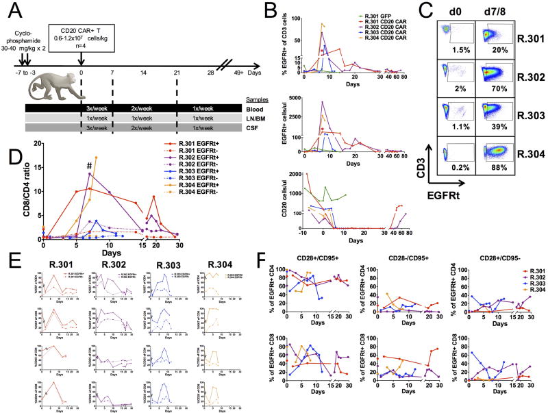
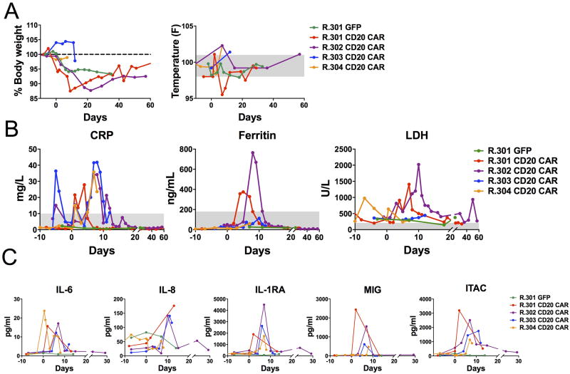
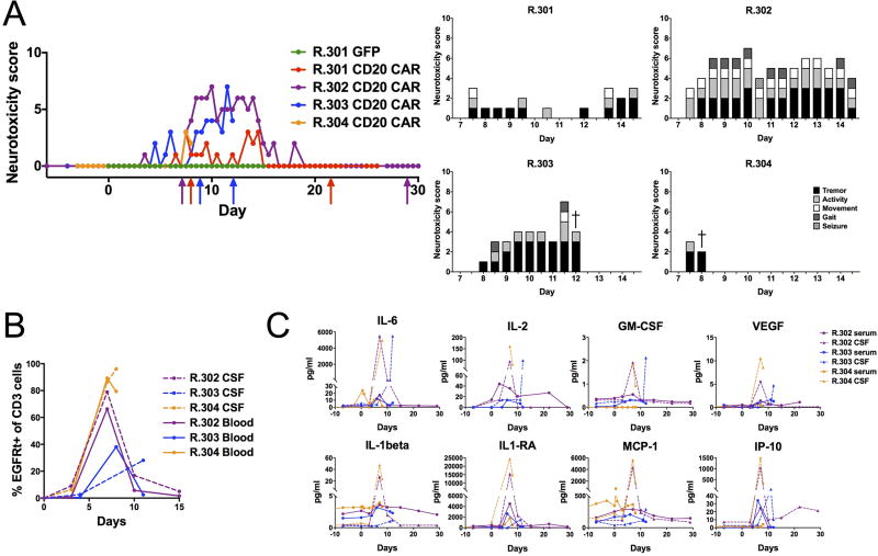
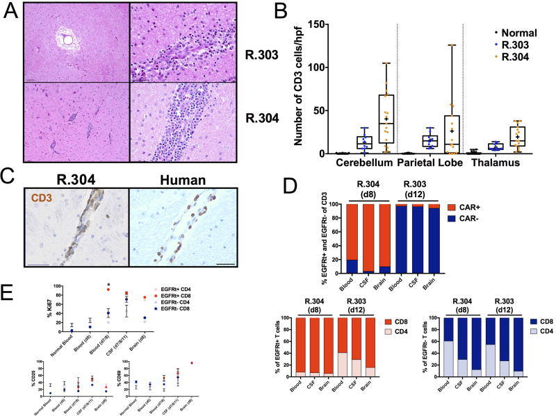
Chimeric antigen receptor (CAR) T-cell immunotherapy has revolutionized the treatment of refractory leukemias and lymphomas, but is associated with significant toxicities, namely cytokine release syndrome (CRS) and neurotoxicity. A major barrier to developing therapeutics to prevent CAR T cell-mediated neurotoxicity is the lack of clinically relevant models. Accordingly, we developed a rhesus macaque (RM) model of neurotoxicity via adoptive transfer of autologous CD20-specific CAR T cells. Following cyclophosphamide lymphodepletion, CD20 CAR T cells expand to 272 to 4,450 cells/μL after 7 to 8 days and elicit CRS and neurotoxicity. Toxicities are associated with elevated serum IL6, IL8, IL1RA, MIG, and I-TAC levels, and disproportionately high cerebrospinal fluid (CSF) IL6, IL2, GM-CSF, and VEGF levels. During neurotoxicity, both CD20 CAR and non-CAR T cells accumulate in the CSF and in the brain parenchyma. This RM model demonstrates that CAR T cell-mediated neurotoxicity is associated with proinflammatory CSF cytokines and a pan-T cell encephalitis.Significance: We provide the first immunologically relevant, nonhuman primate model of B cell-directed CAR T-cell therapy-mediated CRS and neurotoxicity. We demonstrate CAR and non-CAR T-cell infiltration in the CSF and in the brain during neurotoxicity resulting in pan-encephalitis, accompanied by increased levels of proinflammatory cytokines in the CSF. Cancer Discov; 8(6); 750-63. ©2018 AACR.This article is highlighted in the In This Issue feature, p. 663.


Conflict of interest statement

Conflict of interest disclosure statement: No potential conflicts of interest were disclosed by the other authors.

Figures

Figure. 1
Figure. 1. RM GFP and CD20 CAR T cell product composition and function
A, GFP and EGFRt expression in RM T cell products transduced with GFP (R.301 GFP, green) or CD20 CAR (R.301–R.304 EGFRt, red); Mock (blue). CD8 and CD4 composition in RM GFP+ (R.301 GFP+) or EGFRt+ T cell products (R.301–R.304 EGFRt+). B, Composition of T cells in CD20 CAR products in EGFRt+ (top) and EGFRt− (bottom) CD4 and CD8 cells (R.301–R.304, n = 4). CD28+/CD95+: central memory, CD28−/CD95+: effector memory and CD28+/CD95−: naive T cells. Horizontal lines represent the mean. C, CD20 antigen expression in human (K562 and CD20-K562), and in RM (B-LCL1 and B-LCL2) cell lines. Cytolytic activity of RM mock-transduced (dashed lines) and CD20 CAR T cells (solid lines) against 51Cr-labeled targets (K562, CD20-K562, B-LCL1 and B-LCL2). n = 3 replicates per point; representative of four recipients.
Figure 2
Figure 2. Adoptive transfer of RM GFP and CD20 CAR T cells and their dynamics in vivo
A, Animal schema for CD20 CAR RM studies with sample collection frequency. B, Percent (top) and absolute numbers (middle) of RM GFP+ or EGFRt+ (CD20 CAR) T cells and absolute number of CD20 cells (bottom) before and at indicated days after adoptive transfer in the blood. * denotes frozen PBMCs for day 7, R.301, CD20 CAR analysis. C, Frequency (%) of EGFRt+ cells of CD3+ T cells on day 0 (d0, prior to adoptive transfer) and on day of peak expansion, day 7 (R.301, R.302 and R. 304) or day 8 (R.303; (d7/8)) after adoptive transfer in the blood. D, EGFRt+ (solid lines) and EGFRt− (dotted lines) CD8 and CD4 T cell dynamics in the blood, expressed as CD8/CD4 ratio. Peak expansion vs pre-infusion EGFRt+ CD8/CD ratio: # P = 0.03. Dots on day 0 represent infused product samples. E, EGFRt+ (solid lines) and EGFRt− (dotted lines), CD4 (top and third from top panels) and CD8 (second from top and bottom panels) T cell proliferation (Ki67; top two panels), and activation (CD25; bottom two panels) in the blood in vivo. Broken lines after day 0 are used to indicate that the measurements on day 0 represent infused products and subsequent samples represent blood samples. Peak expansion measurements for R.301, R.302 and R.304 were obtained on day 7 and for R.303 on day 8. Data are mean +/− s.e.m. F, EGFRt+ CD4 (top) and CD8 (bottom) T cell subset dynamics in the blood in vivo, represented by CD28+/CD95+ (central memory), CD28−/CD95+ (effector memory) and CD28+/CD95− (naive). Dots on day 0 represent infused products.
Figure 3
Figure 3. Clinical and biochemical markers of cytokine release syndrome (CRS) following adoptive transfer of RM CD20 CAR T cells
A, Changes in body weight and temperature (right). Normal temperature range shaded in gray. B, CRP, Ferritin and LDH levels before and at indicated days after GFP or CD20 CAR T cell transfer. Normal ranges are shaded in gray. C, Serum cytokine levels of IL-6, IL-8, IL-1RA, MIG and ITAC before and at indicated days after GFP or CD20 CAR T cell transfer.
Figure 4
Figure 4. Neurologic toxicity is accompanied by increased T cell infiltration and disproportionately elevated cytokine levels in the CSF following adoptive transfer of RM CD20 CAR T cells
A, Neurotoxicity scores before and at indicated days after GFP or CD20 CAR T cell transfer. Arrows indicate the start and finish of Levetiracetam treatment (red:R.301, violet:R.302, blue:R.303). Neurologic symptoms contributing to the neurotoxicity score in R.301–R.304. B, Frequency (%) of EGFRt+ (CD20 CAR) T cells of CD3+ cells in the CSF (dashed lines) and in the blood (solid lines) following CD20 CAR T cell transfer in R.302–R.304. C, IL-6, IL-2, GM-CSF, VEGF, IL-1beta, IL1-RA, MCP-1 and IP-10 CSF (dashed lines) and serum levels (solid lines) before and at indicated days after CD20 CAR T cell transfer (R.302–R.304, n = 3).
Fig 5
Fig 5. Perivascular T cells and infiltrative encephalitis by EGFRt+ (CD20 CAR) and EGFRt− (non-CAR) T cells following adoptive transfer of RM CD20 CAR T cells
A, H&E brain sections show focal perivascular edema (top left) and focal perivascular inflammation (top right) in R.303 and multifocal perivascular infiltration in the basal ganglia (bottom left) and parietal lobe (bottom right) in H&E sections in R.304. Scale bars, left: 100 µm; right: 50 µm). B, Enumeration of CD3+ T cell infiltration (quantified in 10–15 fields/brain region section at 20× magnification) in the cerebellum, parietal lobe and thalamus in normal RM, in R.303 (blue) and in R.304 (orange). Individual dots represent the number of CD3+ T cells per high power field. Box and whiskers represent the minimum and maximum range, 25th and 75th percentiles and the median range for all quantified fields in one section, + symbols indicate the mean. C, Cerebral perivascular CD3+ T cell infiltration in RM (R.304) and in human brain of a patient with neurotoxicity after CD19 CAR T cell therapy. Scale bars: 50 µm. D, Percent EGFRt+ (CD20 CAR, red) and EGFRt− (non-CAR, blue) T cells and CD8:CD4 proportions in EGFRt+ (bottom left) and EGFRt− (bottom right) compartments in the blood, CSF and brain on day 8 (d8, R.304) and day 12 (d12, R.303). E, Fraction (%) of EGFRt+ or EGFRt− CD4 an CD8 T cells expressing Ki-67, CD25 or CD69 in the blood of normal RM controls (n = 5), on day 0, pre-CD20 CAR infusion (d0, n = 4) and on day 7 or 8 (peak CD20 CAR expansion, (days 7–8, n = 4)), in the CSF (on day 7 (R.302), day 11 (R.303) and day 8 (R.304); days 7–8/11, n = 3) and in the brain (on day 8 (d8) in R.304, n = 1). * P < 0.05.

References

    1. Park JH, Geyer MB, Brentjens RJ. CD19-targeted CAR T-cell therapeutics for hematologic malignancies: interpreting clinical outcomes to date. Blood. 2016;127:3312–20. - PMC - PubMed
    1. Maude SL, Teachey DT, Porter DL, Grupp SA. CD19-targeted chimeric antigen receptor T-cell therapy for acute lymphoblastic leukemia. Blood. 2015;125:4017–23. - PMC - PubMed
    1. Tasian SK, Gardner RA. CD19-redirected chimeric antigen receptor-modified T cells: a promising immunotherapy for children and adults with B-cell acute lymphoblastic leukemia (ALL) Ther Adv Hematol. 2015;6:228–41. - PMC - PubMed
    1. Gardner RA, Finney O, Annesley C, Brakke H, Summers C, Leger K, et al. Intent-to-treat leukemia remission by CD19 CAR T cells of defined formulation and dose in children and young adults. Blood. 2017;129:3322–31. - PMC - PubMed
    1. Lee DW, Gardner R, Porter DL, Louis CU, Ahmed N, Jensen M, et al. Current concepts in the diagnosis and management of cytokine release syndrome. Blood. 2014;124:188–95. - PMC - PubMed

Publication types