. 2018 Jul;55(7):469-478.
doi: 10.1136/jmedgenet-2017-105153. Epub 2018 Mar 21.

Monosomy 18p is a risk factor for facioscapulohumeral dystrophy

Affiliations

Monosomy 18p is a risk factor for facioscapulohumeral dystrophy

Judit Balog et al. J Med Genet. 2018 Jul.

Abstract

Background: 18p deletion syndrome is a rare disorder caused by partial or full monosomy of the short arm of chromosome 18. Clinical symptoms caused by 18p hemizygosity include cognitive impairment, mild facial dysmorphism, strabismus and ptosis. Among other genes, structural maintenance of chromosomes flexible hinge domain containing 1 (SMCHD1) is hemizygous in most patients with 18p deletions. Digenic inheritance of a SMCHD1 mutation and a moderately sized D4Z4 repeat on a facioscapulohumeral muscular dystrophy (FSHD) permissive genetic background of chromosome 4 can cause FSHD type 2 (FSHD2).

Objectives: Since 12% of Caucasian individuals harbour moderately sized D4Z4 repeats on an FSHD permissive background, we tested if people with 18p deletions are at risk of developing FSHD.

Methods: To test our hypothesis we studied different cellular systems originating from individuals with 18p deletions not presenting FSHD2 phenotype for transcriptional and epigenetic characteristics of FSHD at D4Z4. Furthermore, individuals with an idiopathic muscle phenotype and an 18p deletion were subjected to neurological examination.

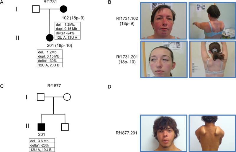
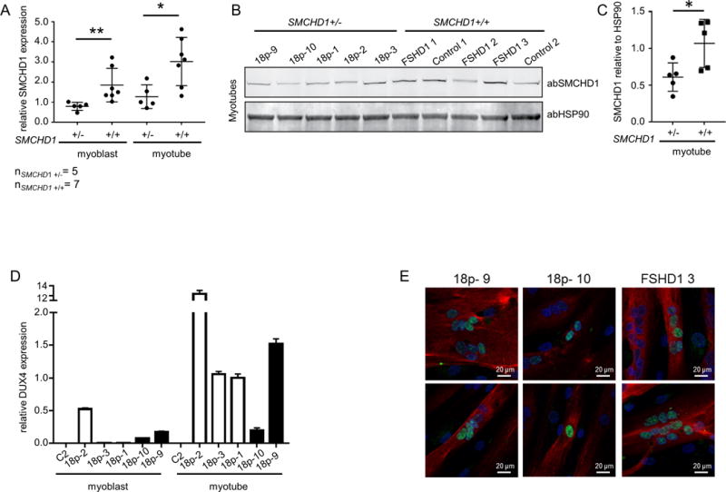
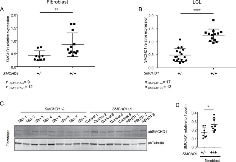
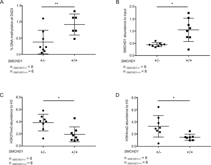
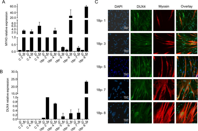
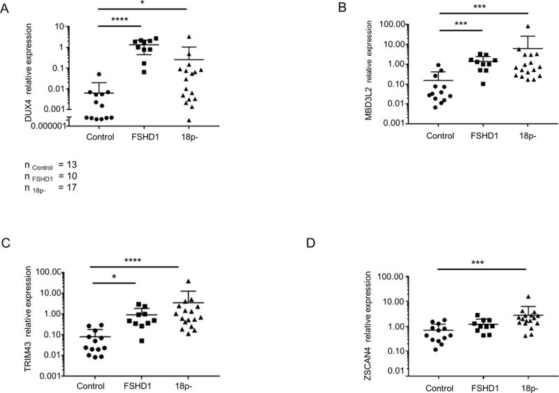
Results: Primary fibroblasts hemizygous for SMCHD1 have a D4Z4 chromatin structure comparable with FSHD2 concomitant with DUX4 expression after transdifferentiation into myocytes. Neurological examination of 18p deletion individuals from two independent families with a moderately sized D4Z4 repeat identified muscle features compatible with FSHD.

Conclusions: 18p deletions leading to haploinsufficiency of SMCHD1, together with a moderately sized FSHD permissive D4Z4 allele, can associate with symptoms and molecular features of FSHD. We propose that patients with 18p deletion should be characterised for their D4Z4 repeat size and haplotype and monitored for clinical features of FSHD.

Keywords: 18p deletion; DUX4; FSHD; SMCHD1; muscle misease.


Conflict of interest statement

Competing interests: None declared.

Figures

Figure 1
Figure 1. Combined 18p- and FSHD phenotype in two independent families
Pedigrees of families Rf1731 (A) and Rf1877 (C). Individuals with a phenotypic combination of 18p- syndrome and FSHD are marked with filled symbols. Below each individual, the size of 18p deletion or duplication is shown, followed by the delta1 values reflecting DNA methylation levels at D4Z4 and the number of D4Z4 units on both chromosomes 4 as well as chromosomal background (A: 4qA FSHD permissive; B: 4qB non-permissive) are depicted. Images of Rf1731.102 and Rf1731.201 (B) and Rf1877.201 (D) highlight facial characteristics compatible with 18p- syndrome, including ocular ptosis, strabismus, low-set ears. Right images show the presence of bilateral winging scapula which are characteristic of FSHD and is more pronounced on Rf1731.102 and Rf1877.201.
Figure 2
Figure 2. Primary myoblasts originating from 18p- individuals presenting with a combined 18p-and FSHD phenotype express DUX4
(A) Relative expression of SMCHD1 normalized to GUSB in control and 18p- myoblasts and myotubes. Values of 18p- 3 were set to 1. (B) Western blot analysis of SMCHD1 and HSP90 in myotubes having one or two copies of SMCHD1. (C) Quantification of western blot signals by normalizing SMCHD1 signal intensities to HSP90 signal intensities. P value was calculated by unpaired t-test followed by Mann-Whitney test. * represents p<0.05 and ** represents p<0.01. (D) Expression levels of DUX4 normalized to GUSB in undifferentiated and differentiated primary myoblast cultures originating from control, from 3 patients with FSHD phenotype and a microdeletion on chromosome 18p without presenting any 18p- phenotype (open bars) and from 18p- 9 and18p- 10 (solid black bars). (E) DUX4 (green signal) immunofluorescence analysis in differentiated myotubes with myosin (red signal) as differentiation marker in primary myotubes originating from 18p- 9, 18p- 10 and FSHD1 3. High content screening was performed using a Nikon confocal microscope at 20× magnification and 2 representative images are shown for every cell culture.
Figure 3
Figure 3
Hemizygosity of SMCHD1 is associated with reduced SMCHD1 transcript and protein levels in different tissues. SMCHD1 transcript levels determined by qRT-PCR and normalized to GUSB in primary fibroblasts (A) and LCLs (B). (C) SMCHD1 protein levels determined by western blot analysis in primary fibroblasts. (D) Intensity of SMCHD1 signal shown on panel C was normalized to Tubulin and statistical analysis of normalized values are shown. Scatter plots show mean and standard error of the mean. Statistical analysis was done by unpaired t-test followed Mann-Whitney test. * represents p<0.05 (D), ** represents p<0.01 (A) and ****represents p<0.0001 (B).
Figure 4
Figure 4. The D4Z4 chromatin landscape is similar in FSHD2 and 18p- syndrome fibroblasts
(A) DNA methylation at D4Z4 in control and 18p- fibroblasts (n=6 control and n=8 18p-) is shown as percentage methylation at the FseI site. (B) SMCHD1 levels at D4Z4 measured by ChIP-qPCR in control and 18p- primary fibroblasts. SMCHD1 enrichment values are shown as normalized to input and enrichment values of control 1 sample were set to 1. H3K27me3 (C) and H3K4me2 (D) levels at D4Z4 in the same samples shown in panel B. Histone modification levels are normalized to H3 and enrichment values of control 1 were set to 1. Statistical analysis was done by unpaired t-test followed Mann-Whitney test. * represents P<0.05 (B, C, D), ** represents P<0.01 (A).
Figure 5
Figure 5
18p- fibroblasts express DUX4 after myogenic transdifferentiation. (A and B) Relative expression of MYH3 and DUX4 in MyoD (M) or GFP (G) transduced control (C) and 18p- primary fibroblasts (18p-). Expression values were normalized to GUSB and value of 18p-2 was set to one. Bar diagrams show the mean and SD of 2 independent experiments. (C) Representative images of DUX4 protein detection by immunofluorescence staining at 20× magnification. Cells were stained with DAPI (nuclei; blue), DUX4 (green), and myogenic differentiation marker MYH1E (red) High content screening was performed by CellomicsArrayscan@VTI HCS instrument at 20× magnification and representative images are shown.
Figure 6
Figure 6
DUX4 and DUX4 target gene expression in control, 18p- and FSHD1 LCLs. Relative expression of DUX4 (A) and DUX4 targets TRIM43 (B), ZSCAN4 (C), and MBD3L2 (D) in a cohort of control, FSHD1 and 18p- LCLs. Scatter plots show expression levels normalized to GUSB and relative expression level of FSHD1–2 was set to 1. Statistical analysis was done by one way ANOVA followed by Kruskal-Wallis test. * represents p<0.05 (A, B), *** represents p<0.01 (C, D) and **** represents p<0.0001 (A, B).

References

    1. Wang LH, Tawil R. Facioscapulohumeral Dystrophy. Current Neurology and Neuroscience Reports. 2016;16(7):66. - PubMed
    1. Lemmers RJ, Tawil R, Petek LM, Balog J, Block GJ, Santen GW, Amell AM, van der Vliet PJ, Almomani R, Straasheijm KR, Krom YD, Klooster R, Sun Y, den Dunnen JT, Helmer Q, Donlin-Smith CM, Padberg GW, van Engelen BG, de Greef JC, Aartsma-Rus AM, Frants RR, de Visser M, Desnuelle C, Sacconi S, Filippova GN, Bakker B, Bamshad MJ, Tapscott SJ, Miller DG, van der Maarel SM. Digenic inheritance of an SMCHD1 mutation and an FSHD-permissive D4Z4 allele causes facioscapulohumeral muscular dystrophy type 2. Nature genetics. 2012;44(12):1370–1374. - PMC - PubMed
    1. Das S, Chadwick BP. Influence of Repressive Histone and DNA Methylation upon D4Z4 Transcription in Non-Myogenic Cells. PloS one. 2016;11(7):e0160022. - PMC - PubMed
    1. Balog J, Thijssen PE, Shadle S, Straasheijm KR, van der Vliet PJ, Krom YD, van den Boogaard ML, de Jong A, RJ FL, Tawil R, Tapscott SJ, van der Maarel SM. Increased DUX4 expression during muscle differentiation correlates with decreased SMCHD1 protein levels at D4Z4. Epigenetics. 2015;10(12):1133–1142. - PMC - PubMed
    1. De Iaco A, Planet E, Coluccio A, Verp S, Duc J, Trono D. DUX-family transcription factors regulate zygotic genome activation in placental mammals. Nature genetics. 2017 - PMC - PubMed

Publication types

MeSH terms

Supplementary concepts