. 2018 Apr 3;115(14):3680-3685.
doi: 10.1073/pnas.1717474115. Epub 2018 Mar 21.

Genetic mapping of species differences via in vitro crosses in mouse embryonic stem cells

Affiliations

Genetic mapping of species differences via in vitro crosses in mouse embryonic stem cells

Stefano Lazzarano et al. Proc Natl Acad Sci U S A. .

Abstract

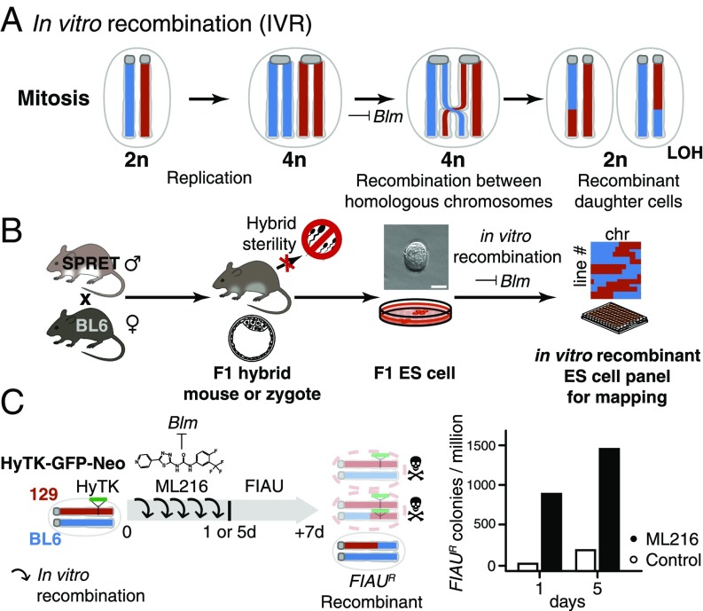
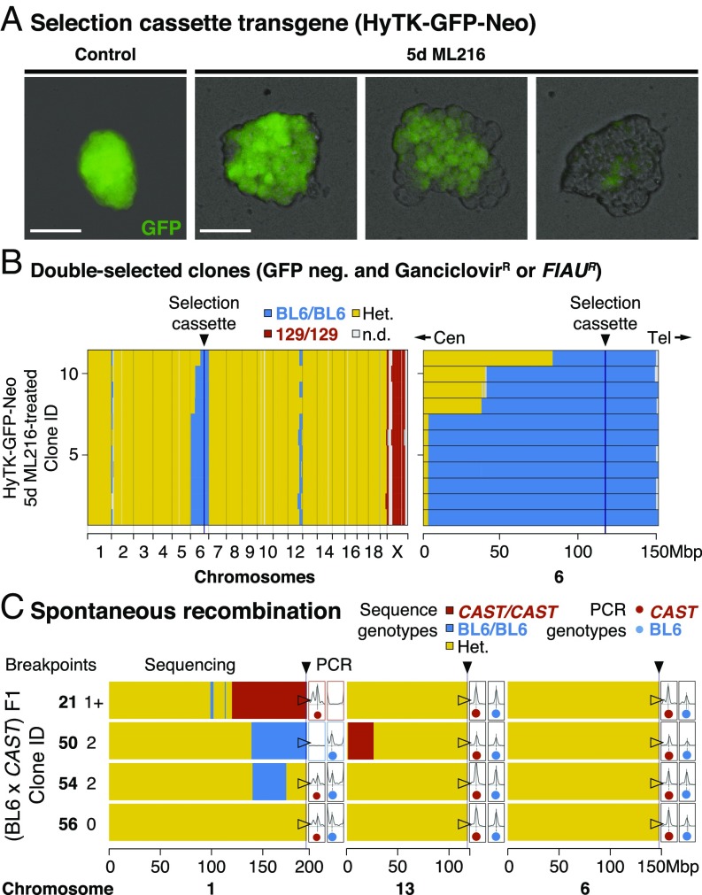
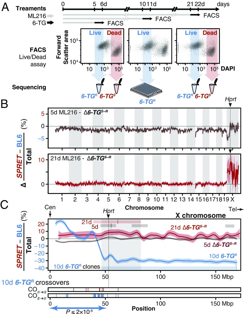
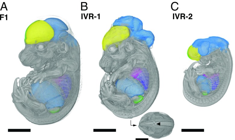
Discovering the genetic changes underlying species differences is a central goal in evolutionary genetics. However, hybrid crosses between species in mammals often suffer from hybrid sterility, greatly complicating genetic mapping of trait variation across species. Here, we describe a simple, robust, and transgene-free technique to generate "in vitro crosses" in hybrid mouse embryonic stem (ES) cells by inducing random mitotic cross-overs with the drug ML216, which inhibits the DNA helicase Bloom syndrome (BLM). Starting with an interspecific F1 hybrid ES cell line between the Mus musculus laboratory mouse and Mus spretus (∼1.5 million years of divergence), we mapped the genetic basis of drug resistance to the antimetabolite tioguanine to a single region containing hypoxanthine-guanine phosphoribosyltransferase (Hprt) in as few as 21 d through "flow mapping" by coupling in vitro crosses with fluorescence-activated cell sorting (FACS). We also show how our platform can enable direct study of developmental variation by rederiving embryos with contribution from the recombinant ES cell lines. We demonstrate how in vitro crosses can overcome major bottlenecks in mouse complex trait genetics and address fundamental questions in evolutionary biology that are otherwise intractable through traditional breeding due to high cost, small litter sizes, and/or hybrid sterility. In doing so, we describe an experimental platform toward studying evolutionary systems biology in mouse and potentially in human and other mammals, including cross-species hybrids.

Keywords: QTL mapping; evolution; genetics; interspecific hybrids; mitotic recombination.


Conflict of interest statement

The authors declare no conflict of interest.

Figures

Fig. 1.
Fig. 1.
IVR via Blm helicase suppression. (A) Blm encodes a helicase normally active during mitosis. Loss of Blm activity leads to increased improper sister chromatid exchange as well as recombination between homologous chromosomes. Mitotic recombination can give rise to recombinant diploid daughter cells with LOH between the breakpoint and the telomeres. (B) IVR allowed the circumvention of hybrid sterility in crosses between the laboratory mouse, e.g., BL6, and a murine sister species SPRET. (BL6 × SPRET)F1 hybrid mice were viable and allowed derivation of F1 ES cells despite male sterility (25). Applying IVR to F1 ES cells allowed rapid and efficient generation of recombinant ES cell panels for genetic mapping. (Scale bar: 50 µm.) chr, chromosome. (C) Efficiency of IVR was estimated by colony survival assay. We estimated the recombination rate between homologous chromosomes with cells hemizygous for a dominant selectable marker (HyTK; green). We induced IVR by adding a small-molecule BLM inhibitor, ML216 (19), to the culturing medium for 1 or 5 d. Under FIAU negative selection, cells having undergone mitotic recombination to become homozygous for the wild-type BL6 alleles (blue) survived, while nonrecombined cells or recombinant cells retaining the HyTK transgene metabolized FIAU, resulting in cell death due to misincorporation of toxic nucleotide analogs (top and middle cells with red chromosomes). Under ML216 treatment (25 µM), IVR rate was estimated to be 2.9 × 10⁻⁴ per cell per generation, yielding 800–1,500 FIAU-resistant colonies per million following treatment.
Fig. 2.
Fig. 2.
Widespread IVR across a range of evolutionary divergence. (A) Selection cassette transgene (HyTK-GFP-Neo). ES cell colonies displayed mosaic GFP expression within a colony when cultured with ML216, but not under control conditions, consistent with homologous recombination and loss of GFP through IVR. Recombination between homologous chromosomes could result in daughter cells with two wild-type (BL6 allele; dark) or transgenic copies (129 allele; bright). Early recombination events followed by random cell loss during clonal expansion could produce completely dark colonies. (Scale bars: 100 µm.) (B) Double-selected clones. After expansion under negative selection against the transgene (both ganciclovir and FIAU kill cells expressing HyTK), 11 ganciclovir-resistant and GFP-negative colonies were whole-genome sequenced. Selection favored loss of transgene (homozygous BL6/BL6 genotypes) at distal chromosome 6. In contrast to normal meiotic recombination (averaging one or more cross-overs per chromosome pair), mitotic recombination typically affected only a single chromosome pair: Much of the genome remained heterozygous (Het.; yellow), with the exception of the transgene-carrying chromosome 6 (mostly BL6/BL6; blue), the single 129 chromosome X (male; 129; red), and at tips of certain chromosomes (e.g., chromosomes 1 and 12). Mitotic recombination events converted genotypes telomeric to the breakpoint toward homozygosity (LOH; yellow to blue). Cen, centromere; n.d., not determined; Tel, telomere. (C) Spontaneous recombination. IVR also occurred in cells carrying divergent genomes with no transgenes. (BL6 × CAST) F1 hybrid ES cells were treated with ML216 and screened by PCR genotyping at diagnostic telomeric markers. Selected clones (two recombinant and control clones each) were whole-genome sequenced, showing recombination events toward both homozygous genotypes, consistent with PCR genotype screening results (total breakpoints per clone ranged from zero to two). Additional recombination events were also recovered, even though the chromosome 1 telomeric marker remained heterozygous (clone 54). These clones also carried nonrecombined chromosomes (e.g., chromosome 6; fully heterozygous; yellow).
Fig. 3.
Fig. 3.
In vitro genetic mapping of variation in 6-TG susceptibility between divergent species. (A) A female ES cell line S18 derived from a M. spretus and BL6 F1 interspecific hybrid was treated with ML216 (25 µM) and subjected to the antimetabolite 6-TG for 1 d before FACS. ES cells were evaluated for viability based on DAPI exclusion. Resistant and susceptible (6-TGR and -TGS) subpopulations were gated conservatively (shaded arrows) and pooled for sequencing. Individual clones from the 10-d ML216 treatment were cultured and whole-genome sequenced. (B) An excess of SPRET contribution on chromosome X between the 6-TGR and -TGS pools suggested that a single locus conferred 6-TG susceptibility. Allele counts were shown as the difference in SPRET bias between the 6-TGs and the 6-TGR samples (as an internal ML216 treatment control) after 5-d (brown) and 21-d (red) ML216 treatment (mean differential SPRET bias ± SEM in megabase windows). In both cases, the genome-wide peak window contains the Hprt gene with the SPRET allele showing significantly increased susceptibility. (C) Detailed view of chromosome X, showing differential SPRET bias for the 5-d (brown) and 21-d (red) ML216 treatment as described above. In addition, 6-TGR clones following 10-d ML216 treatment were sequenced to determine recombination breakpoints. In contrast to the differential bias toward SPRET observed in the susceptible 5- and 21-d ML216 samples, the raw SPRET bias in the solitary 6-TG–resistant sample showed an opposite skew toward BL6 in the 10-d ML216-treatment (blue). Regions deviating from 0 (thus showing bias) after local smoothing in each sample are shown as bars with mark showing maximum skew. Together, they define a common region (shaded) on chromosome X containing Hprt. Cross-overs in individual 6-TGR clones (10-d ML216 treatment) recombined significantly more likely in the SPRET-to-BL6 direction (S > B = 37; B > S = 5; P ≤ 2 × 10⁻⁵) between the centromere (Cen) and Hprt, consistent with strong selection favoring the BL6 Hprtb allele. In contrast, only three additional cross-overs were detected telomeric to Hprt. Tel, telomere.
Fig. 4.
Fig. 4.
Accessing developmental phenotypes in recombinants between evolutionarily divergent species. Embryos at midgestation (14.5 d after fertilization) were derived from nonrecombinant F1 S18 ES cells (A) and IVR lines 1 (B) and 2 (C; see Methods for details). Embryos were dissected, contrast-stained, and scanned by using X-ray microCT at 9.4-µm resolution. The high scanning resolution allowed identification and precise measurements of individual organs (colorized here). Major developmental craniofacial and neural tube closure defects were observed in the IVR lines (B; caudal view with arrowhead indicates neural tube lesion). (Scale bars: 200 µm.)

References

    1. Darwin C. On the Origin of Species by Means of Natural Selection. John Murray; London: 1859.
    1. Dejager L, Libert C, Montagutelli X. Thirty years of Mus spretus: A promising future. Trends Genet. 2009;25:234–241. - PubMed
    1. Lander ES, Botstein D. Mapping Mendelian factors underlying quantitative traits using RFLP linkage maps. Genetics. 1989;121:185–199. - PMC - PubMed
    1. Mackay TFC, Stone EA, Ayroles JF. The genetics of quantitative traits: Challenges and prospects. Nat Rev Genet. 2009;10:565–577. - PubMed
    1. Allen Orr H. The genetics of species differences. Trends Ecol Evol. 2001;16:343–350. - PubMed

Publication types

MeSH terms