

Microglia-Mediated Synapse Loss in Alzheimer's Disease
- PMID: 29563239
- PMCID: PMC6596066
- DOI: 10.1523/JNEUROSCI.1136-17.2017
Microglia-Mediated Synapse Loss in Alzheimer's Disease
Abstract
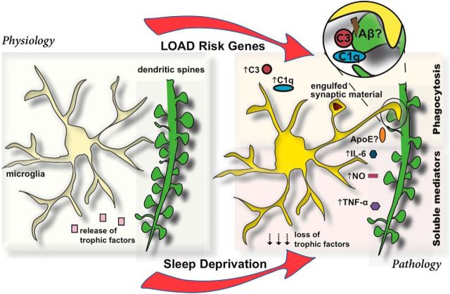
Microglia are emerging as key players in neurodegenerative diseases, such as Alzheimer's disease (AD). Thus far, microglia have rather been known as modulator of neurodegeneration with functions limited to neuroinflammation and release of neurotoxic molecules. However, several recent studies have demonstrated a direct role of microglia in "neuro" degeneration observed in AD by promoting phagocytosis of neuronal, in particular, synaptic structures. While some of the studies address the involvement of the β-amyloid peptides in the process, studies also indicate that this could occur independent of amyloid, further elevating the importance of microglia in AD. Here we review these recent studies and also speculate about the possible cellular mechanisms, and how they could be regulated by risk genes and sleep. Finally, we deliberate on possible avenues for targeting microglia-mediated synapse loss for therapy and prevention.Dual Perspectives Companion Paper: Alzheimer's Disease and Sleep-Wake Disturbances: Amyloid, Astrocytes, and Animal Models by William M. Vanderheyden, Miranda M. Lim, Erik S. Musiek, and Jason R. Gerstner.
Keywords: Alzheimer's disease; amyloid; clearance; microglia; phagocytosis; synaptic pruning.
Copyright © 2018 the authors 0270-6474/18/382911-09$15.00/0.
Figures
References
-
- Achariyar TM, Li B, Peng W, Verghese PB, Shi Y, McConnell E, Benraiss A, Kasper T, Song W, Takano T, Holtzman DM, Nedergaard M, Deane R (2016) Glymphatic distribution of CSF-derived apoE into brain is isoform specific and suppressed during sleep deprivation. Mol Neurodegener 11:74. 10.1186/s13024-016-0138-8 - DOI - PMC - PubMed
-
- Azevedo EP, Ledo JH, Barbosa G, Sobrinho M, Diniz L, Fonseca AC, Gomes F, Romão L, Lima FR, Palhano FL, Ferreira ST, Foguel D (2013) Activated microglia mediate synapse loss and short-term memory deficits in a mouse model of transthyretin-related oculoleptomeningeal amyloidosis. Cell Death Dis 4:e789. 10.1038/cddis.2013.325 - DOI - PMC - PubMed
Publication types
MeSH terms
LinkOut - more resources
Full Text Sources
Other Literature Sources
Medical