. 2018 May 14;92(11):e00303-18.
doi: 10.1128/JVI.00303-18. Print 2018 Jun 1.

Divergent Pathogenic Properties of Circulating Coxsackievirus A6 Associated with Emerging Hand, Foot, and Mouth Disease

Affiliations

Divergent Pathogenic Properties of Circulating Coxsackievirus A6 Associated with Emerging Hand, Foot, and Mouth Disease

Shao-Hua Wang et al. J Virol. .

Abstract

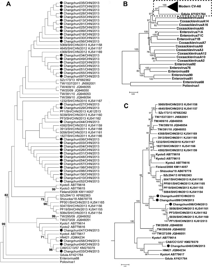
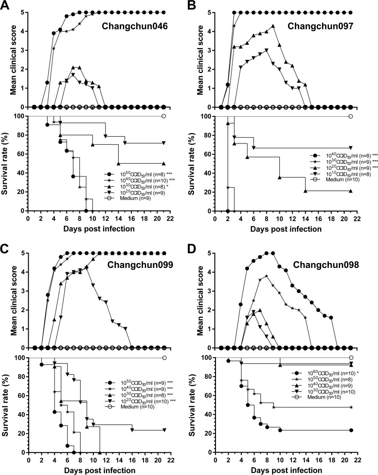
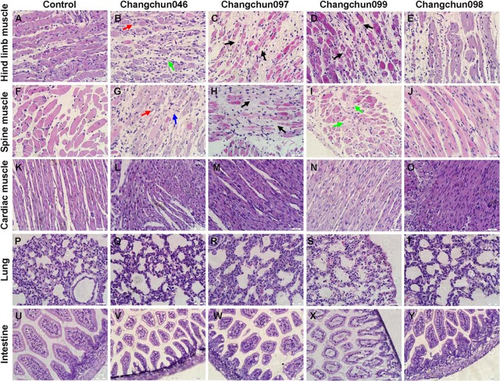
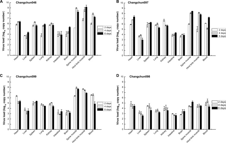
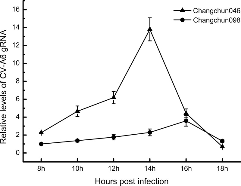
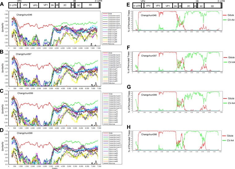
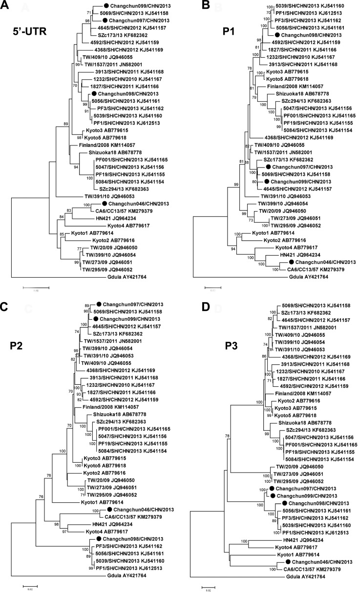
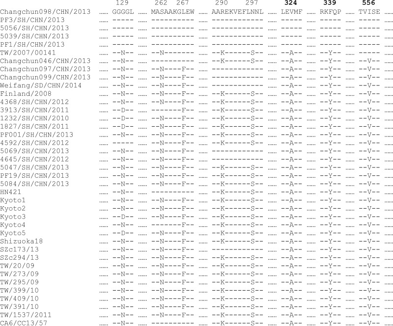
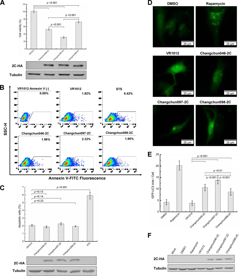
Coxsackievirus A6 (CV-A6) is an emerging pathogen associated with hand, foot, and mouth disease (HFMD). Its genetic characterization and pathogenic properties are largely unknown. Here, we report 39 circulating CV-A6 strains isolated in 2013 from HFMD patients in northeast China. Three major clusters of CV-A6 were identified and related to CV-A6, mostly from Shanghai, indicating that domestic CV-A6 strains were responsible for HFMD emerging in northeast China. Four full-length CV-A6 genomes representing each cluster were sequenced and analyzed further. Bootscanning tests indicated that all four CV-A6-Changchun strains were most likely recombinants between the CV-A6 prototype Gdula and prototype CV-A4 or CV-A4-related viruses, while the recombination pattern was related to, yet distinct from, the strains isolated from other regions of China. Furthermore, different CV-A6 strains showed different capabilities of viral replication, release, and pathogenesis in a mouse model. Further analyses indicated that viral protein 2C contributed to the diverse pathogenic abilities of CV-A6 by causing autophagy and inducing cell death. To our knowledge, this study is the first to report lethal and nonlethal strains of CV-A6 associated with HFMD. The 2C protein region may play a key role in the pathogenicity of CV-A6 strains.IMPORTANCE Hand, foot, and mouth disease (HFMD) is a major and persistent threat to infants and children. Besides the most common pathogens, such as enterovirus A71 (EV-A71) and coxsackievirus A16 (CV-A16), other enteroviruses are increasingly contributing to HFMD. The present study focused on the recently emerged CV-A6 strain. We found that CV-A6 strains isolated in Changchun City in northeast China were associated with domestic origins. These Changchun viruses were novel recombinants of the CV-A6 prototype Gdula and CV-A4. Our results imply that measures to control CV-A6 transmission are urgently needed. Further analyses revealed differing pathogenicities in strains isolated in a neonatal mouse model. One of the possible causes has been narrowed down to the viral protein 2C, using phylogenetic studies, viral sequences, and direct tests on cultured human cells. Thus, the viral 2C protein is a promising target for antiviral drugs to prevent CV-A6-induced tissue damage.

Keywords: and mouth disease; coxsackievirus A6; foot; hand; lethality; mouse model; pathogenicity; recombination.


Figures

FIG 1
FIG 1
Phylogenetic status of modern CV-A6 strains. (A) Neighbor-joining tree based on a short region of the CV-A6 genome (partial VP1, positions 2614 to 2866, corresponding to the Gdula genome). Only topology is shown for clear demonstration of phylogenetic relationships between strains. (B and C) Neighbor-joining tree based on full-length CV-A6 genomes. Prototypes of the HEV-A group, EV-D68, and poliovirus 1 were used as references. The tree indicates that modern CV-A6 strains are phylogenetically close to the prototype Gdula (B), while the subtree indicates that strains from Changchun have different origins (C). All the trees were tested by the bootstrap method for 1,000 replicates, and values of >70 are shown at the nodes. ●, CV-A6-Changchun isolates.
FIG 2
FIG 2
CV-A6-Changchun viruses cause clinical symptoms and mortality in a dose-dependent manner. One-day-old ICR mice (n = 8 to 10 per litter) were intracerebrally inoculated with 10-fold serially diluted dosages of viruses (10 μl/mouse). Control animals were mock infected with MEM instead of virus (10 μl/mouse). Clinical symptoms and mortality were monitored daily for 21 days postinfection. Neonatal mice were challenged with 102.0 to 105.0 CCID50/ml of Changchun046 virus (A), 101.0 to 104.0 CCID50/ml of Changchun097 virus (B), 102.0 to 105.0 CCID50/ml of Changchun099 virus (C), or 103.0 to 106.0 CCID50/ml of Changchun098 virus (D). The log rank test was used to compare the survival rates of newborn mice between the groups and the control group at 21 days postinfection. ***, P < 0.001; *, P < 0.05. One representative of three independent tests is shown.
FIG 3
FIG 3
Pathological analyses of CV-A6-infected newborn mice. One-day-old ICR mice were intracerebrally inoculated with Changchun046 (105.0 CCID50/ml), Changchun097 (104.0 CCID50/ml), Changchun099 (105.0 CCID50/ml), or Changchun098 (105.0 CCID50/ml) or medium (negative control). Infected mice with grade 4 or 5 that exhibited severe hind limb paralysis were dissected for H&E staining. Representative images from the hind limb muscle, spine muscle, cardiac muscle, and lung and intestine tissues after infection are shown. Hind limb muscle (B to D) and spine muscle (G to I) fibers exhibited severe necrosis, including muscle bundle fracture (black arrows), muscle fiber swelling (green arrows), nuclear dissolution (red arrows), and shrinkage (blue arrows), in Changchun046-, Changchun097-, and Changchun099-infected mice. No obvious changes were found in the hind limb muscle and spine muscle fibers of Changchun098-infected mice (E and J), and no detectable pathological changes were observed in the heart (L to O), lung (Q to T), or intestine (V to Y). Magnification, ×400 (scale bars, 20 μm). The results are representative of three independent experiments.
FIG 4
FIG 4
Kinetics of viral load levels in various tissues of CV-A6-infected mice at different time points. One-day-old ICR mice were intracerebrally inoculated with Changchun046 (105.0 CCID50/ml) (A), Changchun097 (104.0 CCID50/ml) (B), Changchun099 (105.0 CCID50/ml) (C), and Changchun098 (105.0 CCID50/ml) (D). The viral loads were detected by qRT-PCR at days 2, 4, and 6 postinfection in 10 samples. The results represent the mean viral loads (log10 copies per milligram of tissue or log10 copies per milliliter of blood) ± SD (3 mice/group; repeated 3 times). The data shown are representative of the results of three independent experiments.
FIG 5
FIG 5
The lethality of CV-A6 correlates with viral replication and release. Shown are one-step growth curves of CV-A6 Changchun046 and Changchun098. Similar amounts of Changchun046 and Changchun098 viruses, based on genome quantification by qRT-PCR, were used to infect RD cells. After adsorbing the virus for 6 h, the infected cells were washed, the medium was replaced with maintenance medium, and the levels of released viruses in the medium were determined through qRT-PCR at 2-h intervals, starting 8 h postadsorption. The values are shown as means ± SD.
FIG 6
FIG 6
Similarity tests and bootscanning analyses of the CV-A6-Changchun strains and the prototype Gdula and CV-A4 on the basis of full-length genomes. The similarity tests for Changchun046 (A), Changchun097 (C), Changchun099 (E), and Changchun098 (G) were performed with prototypic strains of members of the HEV-A group. The test results indicated that modern CV-A6 strains circulating in Changchun were mostly recombinants between CV-A6 and CV-A4, as previously described (16, 17). Thus, for better demonstration, only CV-A4 (accession number AY421762) and CV-A6 Gdula (accession number AY421764) were included as the reference sequences for the bootscanning tests for Changchun046 (B), Changchun097 (D), Changchun099 (F), and Changchun098 (H). EV-D68 (accession number AY426531) and poliovirus 1 (accession number V01150) were used as the outliers in both similarity tests and bootscanning analyses. All the tests were performed with full-length viral genomes. A cartoon of the enteroviral genome is shown at the top.
FIG 7
FIG 7
Phylogenetic trees constructed for 5′ UTR, P1, P2, and P3 regions of CV-A6-Changchun strains with other CV-A6 strains retrieved from the GenBank database. Neighbor-joining trees based on the full-length 5′ UTR (A), P1 (B), P2 (C), and P3 (D) regions were generated. All the trees were tested by bootstrapping for 1,000 replicates, and values of >70 are shown at the nodes. ●, CV-A6-Changchun isolates.
FIG 8
FIG 8
Sequence alignment of the lethal and nonlethal strains of CV-A6. The amino acid sequences of the P2 regions of CV-A6 strains from Changchun and other places in China and worldwide were aligned, and specific positions possibly associated with lethality are indicated, based on the differences between the lethal group (containing Changchun046, Changchun097, and Changchun099) and the nonlethal group (containing Changchun098). Dashes stand for amino acid residues identical to those of Changchun098.
FIG 9
FIG 9
CV-A6 2C protein induces cell death through autophagy instead of apoptosis in RD cells. (A) Cell lethality caused by different CV-A6 2C proteins in RD cells. Changchun046 2C-, Changchun097 2C-, or Changchun098 2C- expressing vectors were transfected into RD cells, and a cell proliferation assay was performed at 48 h posttransfection. Western blotting results indicating expression levels of viral 2C proteins for the cell lethality experiment are shown. (B) Representative flow charts for the apoptosis analysis. Changchun046 2C-, Changchun097 2C-, or Changchun098 2C-expressing vectors were transfected into RD cells. The RD cells were harvested at 48 h posttransfection, and cell death was evaluated by flow cytometry. The percentages of apoptotic cells corresponding to the increased FITC fluorescence intensity for each of the experimental conditions are indicated. RD cells transfected with VR1012 or treated with STS were used as negative and positive controls, respectively. (C) Bar graph of apoptosis analysis in RD cells. Shown are Western blotting results indicating the expression levels of viral 2C proteins from panel B. (D) Live-cell imaging showing GFP-LC3 aggregation. RD cells were cotransfected with pEGFP-LC3 and the VR1012 empty vector, Changchun046 2C, Changchun097 2C, or Changchun099 2C. Rapamycin was used as a positive control to induce autophagy. The GFP-LC3 aggregations in the cells were observed by fluorescence microscopy at 48 h posttransfection. Representative images are shown. Magnification, ×400. (E) Bar graph indicating average GFP-LC3 dot formation in RD cells expressing CV-A6 2C. Each bar value indicates the average number of GFP dots in 10 cells per sample. Rapamycin was used as a positive control. (F) Western blotting results showing 2C protein expression in RD cells from panel E. The values in panels A, C, and E are shown as means ± SD.

References

    1. Bendig JW, Fleming DM. 1996. Epidemiological, virological, and clinical features of an epidemic of hand, foot, and mouth disease in England and Wales. Commun Dis Rep CDR Rev 6:R81–R86. - PubMed
    1. Chan KP, Goh KT, Chong CY, Teo ES, Lau G, Ling AE. 2003. Epidemic hand, foot and mouth disease caused by human enterovirus 71, Singapore. Emerg Infect Dis 9:78–85. doi: 10.3201/eid1301.020112. - DOI - PMC - PubMed
    1. Shimizu H, Utama A, Onnimala N, Li C, Li-Bi Z, Yu-Jie M, Pongsuwanna Y, Miyamura T. 2004. Molecular epidemiology of enterovirus 71 infection in the Western Pacific region. Pediatr Int 46:231–235. doi: 10.1046/j.1442-200x.2004.01868.x. - DOI - PubMed
    1. Ho M, Chen ER, Hsu KH, Twu SJ, Chen KT, Tsai SF, Wang JR, Shih SR. 1999. An epidemic of enterovirus 71 infection in Taiwan. Taiwan Enterovirus Epidemic Working Group. N Engl J Med 341:929–935. - PubMed
    1. Brown BA, Oberste MS, Alexander JP Jr, Kennett ML, Pallansch MA. 1999. Molecular epidemiology and evolution of enterovirus 71 strains isolated from 1970 to 1998. J Virol 73:9969–9975. - PMC - PubMed

Publication types

LinkOut - more resources