. 2018 Mar 22;3(6):e94463.
doi: 10.1172/jci.insight.94463.

Pro-resolving mediators promote resolution in a human skin model of UV-killed Escherichia coli-driven acute inflammation

Affiliations

Pro-resolving mediators promote resolution in a human skin model of UV-killed Escherichia coli-driven acute inflammation

Madhur P Motwani et al. JCI Insight. .

Abstract

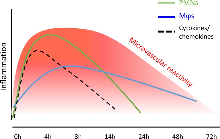
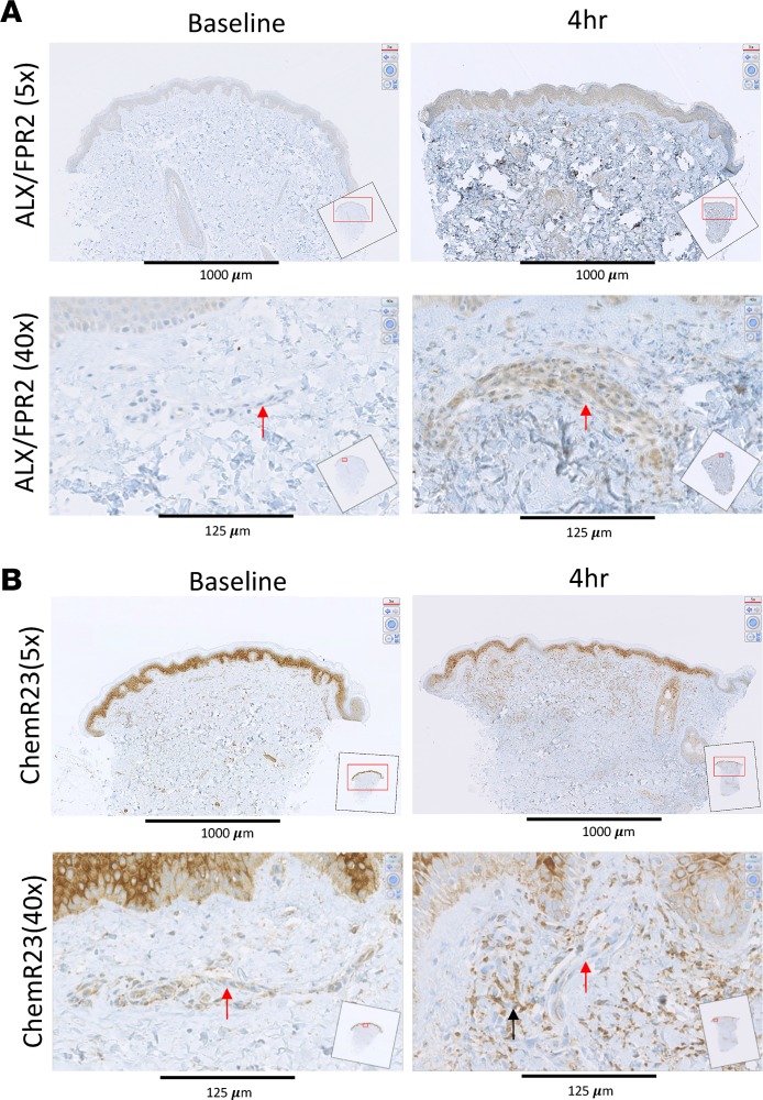
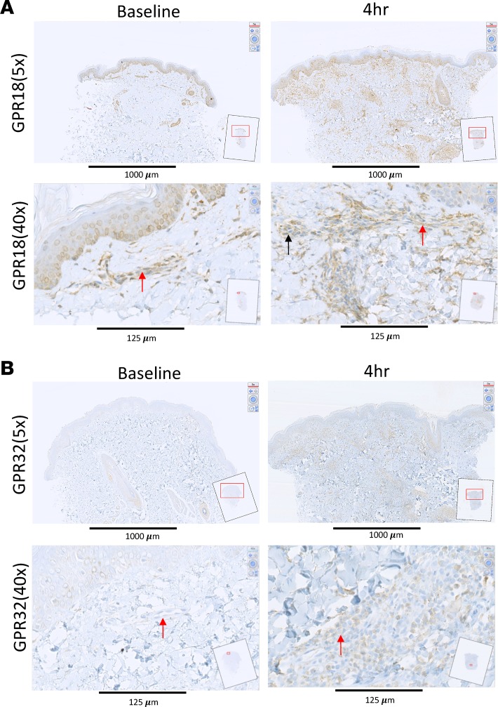
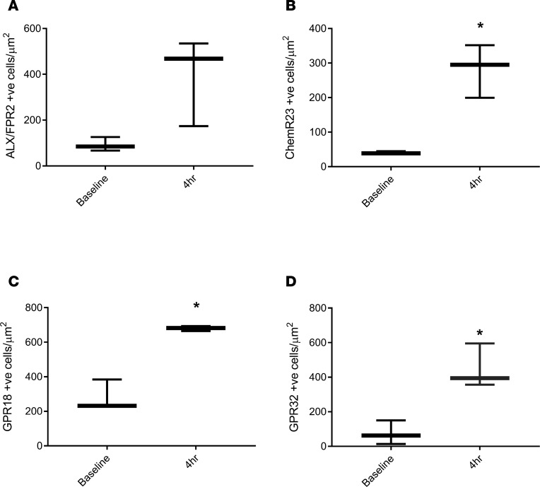
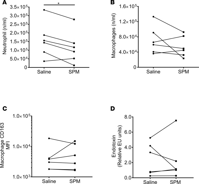
While the treatment of inflammatory disorders is generally based on inhibiting factors that drive onset of inflammation, these therapies can compromise healing (NSAIDs) or dampen immunity against infections (biologics). In search of new antiinflammatories, efforts have focused on harnessing endogenous pathways that drive resolution of inflammation for therapeutic gain. Identification of specialized pro-resolving mediators (SPMs) (lipoxins, resolvins, protectins, maresins) as effector molecules of resolution has shown promise in this regard. However, their action on inflammatory resolution in humans is unknown. Here, we demonstrate using a model of UV-killed Escherichia coli-triggered skin inflammation that SPMs are biosynthesized at the local site at the start of resolution, coinciding with the expression of receptors that transduce their actions. These include receptors for lipoxin A4 (ALX/FPR2), resolvin E1 (ChemR23), resolvin D2 (GPR18), and resolvin D1 (GPR32) that were differentially expressed on the endothelium and infiltrating leukocytes. Administering SPMs into the inflamed site 4 hours after bacterial injection caused a reduction in PMN numbers over the ensuing 6 hours, the phase of active resolution in this model. These results indicate that in humans, the appearance of SPMs and their receptors is associated with the beginning of inflammatory resolution and that their therapeutic supplementation enhanced the resolution response.

Keywords: Eicosanoids; Inflammation.


Conflict of interest statement

Conflict of interest: The authors have declared that no conflict of interest exists.

Figures

Figure 1
Figure 1. Summary of resolution markers in the UVkEc-triggered self-limiting acute inflammation model.
Acute inflammation was triggered in the forearm of healthy volunteers by intradermal injection of 1.5 × 10⁷ UV-killed E. coli (UVkEc). Inflammatory exudate at the injection site was acquired into a suction blister. Temporal profile of inflammatory mediators as well as leukocytes in the exudate is depicted as published previously (23). MΦs, macrophages.
Figure 2
Figure 2. Human skin blister bioactive LM-SPM signature profiles.
Acute inflammation was triggered in the forearm of healthy volunteers by intradermal injection of 1.5 × 10⁷ UV-killed E. coli (UVkEc). Inflammatory exudate at the injection site was acquired into a suction blister raised at 0, 4, 8, 14, and 24 hours. Infiltrated cells were centrifuged and cell-free supernatants were analyzed by lipid mediator (LM) metabololipidomics. (A) Representative multiple reaction monitoring (MRM) chromatograms for identified mediators in skin blister exudates. (B) Characteristic MS/MS fragmentation spectra employed in the identification of RvD1, LXA4, and MaR1. Results are representative of n = 34 exudates. Table 1 displays LM-SPM concentrations for the identified mediators. n = 6–7 donors per time point. Supplemental Figure 1 displays MS/MS spectra employed for the identification of mediators from the arachidonic acid, docosahexaenoic acid, and eicosapentaenoic acid bioactive metabolomes. SPM, specialized pro-resolving mediator.
Figure 3
Figure 3. Temporal shift in LM-SPM concentrations in human skin blister exudates during inflammation-resolution.
Acute inflammation was triggered in the forearm of healthy volunteers by intradermal injection of 1.5 × 10⁷ UV-killed E. coli (UVkEc). Inflammatory exudate at the injection site was acquired into a suction blister raised at 0, 4, 8, 14, and 24 hours. Cell-free exudates were obtained and lipid mediator identity and concentrations determined as in Figure 2. (A) Partial least squares-discriminant analysis 2-dimensional score plots of human skin blister LM-SPM exudates at 0, 4, 8, 14, and 24 hours. Gray ellipse denotes 95% confidence region. (B) 2-Dimensional score plot depicting the contribution of distinct mediators to the score plots. Concentrations for (C) PGE2, (D) 20-OH-LTB4, (E) LXB4, (F) RvE3, (G) LXA4, and (H) MaR1 at distinct intervals following UVkEc injection. Statistical comparison between groups was assessed by performing Kruskal-Wallis test and a post hoc Dunnet’s test to correct for multiple comparisons. n = 6–7 volunteers per interval. *P < 0.05 and ***P < 0.001 versus 0-hour time point; #P < 0.05 versus 8-hour mediator levels. LM-SPM, lipid mediator/specialized pro-resolving mediator.
Figure 4
Figure 4. SPM receptors are differentially expressed on the endothelium and the infiltrating leukocytes — ALX/FPR2 and ChemR23.
Acute inflammation was triggered in the ventral aspect of forearm of healthy volunteers by the intradermal injection of 1.5 × 10⁷ UV-killed E. coli (UVkEc) suspended in 100 μl of sterile saline. Four hours after injection, a 3-mm skin punch biopsy was taken from the inflamed site under local anesthesia. Naive skin was treated as baseline. Formalin-fixed paraffin-embedded skin sections were probed by immunohistochemistry for receptor identification. Low-magnification (×5) and high-magnification (×40) images at baseline and at the 4-hour time point are shown here for ALX/FPR2 (A) and ChemR23 (B). Red arrows highlight the endothelium and black arrow highlights the infiltrating leukocytes. Representative images from n = 3. SPM, specialized pro-resolving mediator.
Figure 5
Figure 5. SPM receptors are differentially expressed on the endothelium and the infiltrating leukocytes — GPR18 and GPR32.
Acute inflammation was triggered in the ventral aspect of forearms of healthy volunteers by the intradermal injection of 1.5 × 10⁷ UV-killed E. coli (UVkEc) suspended in 100 μl of sterile saline. Four hours after injection, a 3-mm skin punch biopsy was taken from the inflamed site under local anaesthesia. Naive skin was treated as baseline. Formalin-fixed paraffin-embedded skin sections were probed by immunohistochemistry for receptor identification. Low-magnification (×5) and high-magnification (×40) images at baseline and at the 4-hour time point are shown here for GPR18 (A) and GPR32 (B). Red arrows highlight the endothelium and the black arrow highlights the infiltrating leukocytes. Representative images from (n = 3). SPM, specialized pro-resolving mediator.
Figure 6
Figure 6. SPM receptor expression increases during acute inflammation.
Acute inflammation was triggered in the ventral aspect of forearms of healthy volunteers by the intradermal injection of 1.5 × 10⁷ UV-killed E. coli (UVkEc) suspended in 100 μl of sterile saline. Four hours after injection, a 3-mm skin punch biopsy was taken from the inflamed site under local anaesthesia. Formalin-fixed paraffin-embedded skin sections were probed by immunohistochemistry for receptor identification, as shown in Figures 4 and 5. Increase in receptor expression from baseline to 4 hours following UVkEc injection for ALX/FPR2 (A), ChemR23 (B), GPR18 (C), and GPR32 (D) is shown here. Data expressed as box-and-whisker plots (box representing the median and the whiskers representing the maximum-minimum values). Statistical comparison between baseline and 4-hour expression was assessed by Wilcoxon’s matched-pairs test. n = 3 for each time point. *P < 0.05. SPM, specialized pro-resolving mediator.
Figure 7
Figure 7. SPMs enhance neutrophil clearance.
Specialized pro-resolving mediators (SPMs) (570 pM LXB4, 115 pM RvE1, 450 pM RvD2, 3.2 nM AT-RvD1 in 100 μl saline) were injected into the established inflamed site 4 hours after UV-killed E. coli (UVkEc), with an equivalent volume of saline injected into the contralateral forearm. Six hours later (or 10 hours after bacterial injection), a negative-pressure suction cup was placed over the inflamed site to acquire total inflammatory cells. Actions of SPMs on (A) PMN numbers were determined by polychromatic flow cytometry. Polychromatic flow cytometry was also used to determine the influence of SPMs on (B) macrophage numbers as well as (C) macrophage CD163 expression levels. Actions of SPMs on (D) endotoxin clearance 10 hours after UVkEc injection. Statistical comparison between groups was assessed by 2-tailed paired Student’s t test. Data are expressed as a before/after dot plot, n = 6/group. *P < 0.05.

Similar articles

Cited by

References

    1. Segal AW, Peters TJ. Characterisation of the enzyme defect in chronic granulomatous disease. Lancet. 1976;1(7974):1363–1365. - PubMed
    1. Morgenstern DE, Gifford MA, Li LL, Doerschuk CM, Dinauer MC. Absence of respiratory burst in X-linked chronic granulomatous disease mice leads to abnormalities in both host defense and inflammatory response to Aspergillus fumigatus. J Exp Med. 1997;185(2):207–218. doi: 10.1084/jem.185.2.207. - DOI - PMC - PubMed
    1. Franceschi C, Campisi J. Chronic inflammation (inflammaging) and its potential contribution to age-associated diseases. J Gerontol A Biol Sci Med Sci. 2014;69 Suppl 1:S4–S9. - PubMed
    1. Sfriso P, et al. Infections and autoimmunity: the multifaceted relationship. J Leukoc Biol. 2010;87(3):385–395. doi: 10.1189/jlb.0709517. - DOI - PubMed
    1. Vane JR, Botting RM. Anti-inflammatory drugs and their mechanism of action. Inflamm Res. 1998;47 Suppl 2:S78–S87. - PubMed

Publication types

MeSH terms

Substances