. 2018 Mar 22;3(6):e96272.
doi: 10.1172/jci.insight.96272.

Endothelial cell Pannexin1 modulates severity of ischemic stroke by regulating cerebral inflammation and myogenic tone

Affiliations

Endothelial cell Pannexin1 modulates severity of ischemic stroke by regulating cerebral inflammation and myogenic tone

Miranda E Good et al. JCI Insight. .

Abstract

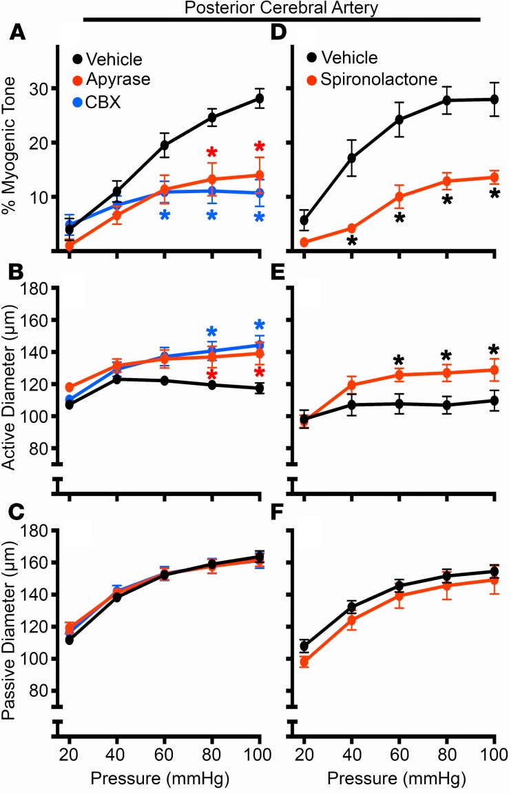
Ischemic stroke is a leading cause of morbidity and mortality in the US; however, there currently exists only one effective acute pharmacological therapeutic intervention. Purinergic signaling has been shown to regulate vascular function and pathological processes, including inflammation and arterial myogenic reactivity, and plays a role in ischemic stroke outcome. Purinergic signaling requires extracellular purines; however, the mechanism of purine release from cells is unclear. Pannexin1 (Panx1) channels are potentially novel purine release channels expressed throughout the vascular tree that couples regulated purine release with purinergic signaling. Therefore, we examined the role of smooth muscle and endothelial cell Panx1, using conditional cell type-specific transgenic mice, in cerebral ischemia/reperfusion injury outcomes. Deletion of endothelial cell Panx1, but not smooth muscle cell Panx1, significantly reduced cerebral infarct volume after ischemia/reperfusion. Infiltration of leukocytes into brain tissue and development of cerebral myogenic tone were both significantly reduced when mice lacked endothelial Panx1. Panx1 regulation of myogenic tone was unique to the cerebral circulation, as mesenteric myogenic reactivity and blood pressure were independent of endothelial Panx1. Overall, deletion of endothelial Panx1 mitigated cerebral ischemic injury by reducing inflammation and myogenic tone development, indicating that endothelial Panx1 is a possible novel target for therapeutic intervention of ischemic stroke.

Keywords: Mouse models; Neuroscience; Stroke.


Conflict of interest statement

Conflict of interest: The authors have declared that no conflict of interest exists.

Figures

Figure 1
Figure 1. Panx1 is expressed in EC and SMC of cerebral arteries with successful knockdown in EC and SMC transgenic mice.
In all sections, Panx1 (Panx1-CT antibody) was stained in red and nuclei (DAPI) in blue. (A) Low-magnification view of brain section. Arrow indicates cerebral artery in field of view. (B) Zoomed-in cross section of the somatosensory cortex neurons sections were stained for Panx1 and neurons using NeuN antibody (green). (C) Isolated control PCA sections were collected from Panx1 fl/fl (control) at low magnification, as well as (D) secondary antibody only and (E) rabbit IgG. (F) High magnification of control PCA and PCA from mice that had genotypes of SMC Panx1 Δ/Δ (G), and EC Panx1 Δ/Δ (H). In D and E, the autofluorescence of the internal elastic lamina is in green and separates the EC (E) and SMC (S) layers. In FH the separation between EC and SMC, the IEL, is indicated by a dotted line. Asterisks indicate lumen of artery in each image. (A and B) Scale bars: 100 μm. (C–E) Scale bar: 20 μm. (F–H) Scale bar: 10 μm.
Figure 2
Figure 2. Deletion of EC Panx1 significantly reduces stroke infarct volume.
Brains were isolated from mice following a 90-minute MCA occlusion and 24-hour reperfusion and stained with TTC to quantify infarct volume. Representative brains, in 2-mm sections, are shown for SMC Panx fl/fl (32.3% infarct, A), SMC Panx Δ/Δ mice (37.5% infarct, B), EC Panx1 fl/fl (38.9% infarct, D), EC Panx1 Δ/Δ mice (8.7% infarct, E), and Cre Panx1 fl/fl (33.6% infarct, F). Quantification of infarct volume is shown for each genotype (C and G). SMC Panx1 fl/fl, n = 9; SMC Panx1 Δ/Δ, n = 11; EC Panx1 fl/fl, n = 9; EC Panx1 Δ/Δ, n = 9; Cre Panx1fl/fl, n = 6 mice. *P < 0.05; 1-way ANOVA/Tukey’s multiple comparisons post hoc test vs. EC Panx1 fl/fl (P = 0.09 for EC Panx1 Δ/Δ vs. Cre Panx1fl/fl).
Figure 3
Figure 3. Brain tissue infiltration of leukocytes is significantly reduced in EC Panx1 Δ/Δ mice following stroke.
Following a 90-minute MCAO and 24-hour reperfusion, ischemic and contralateral hemispheres were perfused and isolated from EC Panx1 fl/fl and EC Panx1 Δ/Δ mice and stained for leukocyte markers for flow cytometry analysis. Differences in the percentages of total infiltrated leukocytes (CD45hi), neutrophils (CD45hi/Ly6G+), or monocytes (CD45hi/CD11b+/Ly6C+) between the ischemic and contralateral hemispheres were calculated (A–C). Representative flow cytometry graphs of the contralateral (D) and ischemic (E) hemispheres of an EC Panx1 fl/fl mouse are shown, along with gating strategies. Live cells (red box) were further analyzed for expression of CD45. Intermediate expression of CD45 is indicative of microglia cells (black box), while cells expressing high levels of CD45 (green box), indicative of infiltrated leukocytes, were further subdivided into Ly6G-positive or CD11b and Ly6C double-positive cells. EC Panx1 fl/fl, n = 6 mice; EC Panx1 Δ/Δ, n = 5 mice. *P < 0.05; Student’s t test.
Figure 4
Figure 4. Degradation of extracellular nucleotides or inhibition of Panx1 channels significantly blunts development of cerebral myogenic tone.
Isolated PCAs from C57Bl/6 control mice were pressurized, and intraluminal pressure was incrementally increased from 20–100 mmHg in the presence or absence of apyrase (10 U/ml; A–C), carbenoxolone (CBX; 50 μM; A–C), or spironolactone (80 μM; D–F). Percent myogenic tone (A and D), active diameter (Ca2+ present in solution; B and E), and passive diameter (Ca2+-free solution; C and F) were evaluated over the range of pressures. For each group in A–C, n = 6 mice; Spironolactone, n = 4; and the vehicle control, n = 7 mice. *P < 0.05 vs. vehicle control. Two-way ANOVA/Sidak’s multiple comparisons post hoc test.
Figure 5
Figure 5. Development of myogenic tone in cerebral arteries is dependent upon EC Panx1 but not SMC Panx1.
Percent myogenic tone, active diameter, and passive diameter of isolated, pressurized PCAs from EC Panx1 (fl/fl and Δ/Δ; A–C) and SMC Panx1 (fl/fl and Δ/Δ; D–F) mice were analyzed. EC Panx1 fl/fl, n = 6; EC Panx1 Δ/Δ, n = 8; SMC Panx1 fl/fl, n = 4; and SMC Panx1 Δ/Δ, n = 6 mice. *P < 0.05. Two-way ANOVA/Sidak’s multiple comparisons post hoc test.
Figure 6
Figure 6. Myogenic tone in mesenteric arteries, mean arterial pressure, and heart rate are independent of EC Panx1.
Pressure curves were run on third-order mesenteric arteries isolated from C57Bl/6 mice in the presence or absence of carbenoxolone (CBX; 50 μM; A–C), spironolactone (80 μM; D–F), or mesenteric arteries isolated from EC Panx1 (fl/fl and Δ/Δ) mice (G–I). Percent myogenic tone, active diameter, and passive diameter were analyzed for each group. Control (CBX), n = 5; CBX, n = 3; control (spironolactone), n = 3; spironolactone, n = 3; EC Panx1 fl/fl, n = 4; and EC Panx1 Δ/Δ, n = 6 mice. *P < 0.05. Two-way ANOVA/Sidak’s multiple comparisons post hoc test. Telemetry was used to collect average day and night mean arterial pressure (J) and heart rate (K) from EC Panx1 fl/fl and EC Panx1 Δ/Δ mice. No significant differences were found using Student’s t test.

Similar articles

Cited by

References

    1. Go AS, et al. Heart disease and stroke statistics--2014 update: a report from the American Heart Association. Circulation. 2014;129(3):e28–e292. doi: 10.1161/01.cir.0000441139.02102.80. - DOI - PMC - PubMed
    1. Fugate JE, Rabinstein AA. Update on intravenous recombinant tissue plasminogen activator for acute ischemic stroke. Mayo Clin Proc. 2014;89(7):960–972. doi: 10.1016/j.mayocp.2014.03.001. - DOI - PubMed
    1. Bond SR, Naus CC. The pannexins: past and present. Front Physiol. 2014;5:58. - PMC - PubMed
    1. Lohman AW, et al. Expression of pannexin isoforms in the systemic murine arterial network. J Vasc Res. 2012;49(5):405–416. doi: 10.1159/000338758. - DOI - PMC - PubMed
    1. Burns AR, Phillips SC, Sokoya EM. Pannexin protein expression in the rat middle cerebral artery. J Vasc Res. 2012;49(2):101–110. doi: 10.1159/000332329. - DOI - PMC - PubMed

Publication types

MeSH terms