. 2018 Mar 22;3(6):e98459.
doi: 10.1172/jci.insight.98459.

Enzymatically oxidized phospholipids restore thrombin generation in coagulation factor deficiencies

Affiliations

Enzymatically oxidized phospholipids restore thrombin generation in coagulation factor deficiencies

David A Slatter et al. JCI Insight. .

Abstract

Hemostatic defects are treated using coagulation factors; however, clot formation also requires a procoagulant phospholipid (PL) surface. Here, we show that innate immune cell-derived enzymatically oxidized phospholipids (eoxPL) termed hydroxyeicosatetraenoic acid-phospholipids (HETE-PLs) restore hemostasis in human and murine conditions of pathological bleeding. HETE-PLs abolished blood loss in murine hemophilia A and enhanced coagulation in factor VIII- (FVIII-), FIX-, and FX-deficient human plasma . HETE-PLs were decreased in platelets from patients after cardiopulmonary bypass (CPB). To explore molecular mechanisms, the ability of eoxPL to stimulate individual isolated coagulation factor/cofactor complexes was tested in vitro. Extrinsic tenase (FVIIa/tissue factor [TF]), intrinsic tenase (FVIIIa/FIXa), and prothrombinase (FVa/FXa) all were enhanced by both HETE-PEs and HETE-PCs, suggesting a common mechanism involving the fatty acid moiety. In plasma, 9-, 15-, and 12-HETE-PLs were more effective than 5-, 11-, or 8-HETE-PLs, indicating positional isomer specificity. Coagulation was enhanced at lower lipid/factor ratios, consistent with a more concentrated area for protein binding. Surface plasmon resonance confirmed binding of FII and FX to HETE-PEs. HETE-PEs increased membrane curvature and thickness, but not surface charge or homogeneity, possibly suggesting increased accessibility to cations/factors. In summary, innate immune-derived eoxPL enhance calcium-dependent coagulation factor function, and their potential utility in bleeding disorders is proposed.

Keywords: Coagulation; Hematology.


Conflict of interest statement

Conflict of interest: The authors have declared that no conflict of interest exists.

Figures

Figure 1
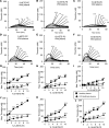
Figure 1. Hydroxyeicosatetraenoic acid–phospholipids (HETE-PLs) restore thrombin generation in FVIII-deficient plasma.
Thrombin generation was measured using thrombinoscope as indicated in Methods using either healthy pooled or FVIII-deficient plasma. (A–F) Representative traces showing effect of HETE-PL positional isomers on thrombin generation in FVIII-deficient plasma. These were repeated 3 times to generate data shown in G–L. (G–L) Summary data showing the effect of dose dependence of HETE-PLs on coagulation (n = 3, mean ± SEM). Square, normal plasma; triangle, FVIII deficient plasma, **P < 0.01, *P < 0.05 compared with no HETE-PL, as determined by 1-way ANOVA and post hoc Tukey tests.
Figure 2
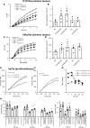
Figure 2. HETE-PLs enhance thrombin generation in the absence of FIX or FXI, or in the absence of tissue factor pathway inhibitor (TFPI) and FVIII combined.
(A and B) Thrombin generation was initiated by addition of liposomes to plasma deficient in FIX, as described in Methods. Liposomes were generated as described in Methods, and 10% 1-stearoyl-2-arachidonyl-PE (SAPE) was replaced with 10% 15-HETE-PE where indicated. (C and D) Thrombin generation was initiated by addition of liposomes to plasma deficient in FXI, as described in Methods. Liposomes were generated as described in Methods, and 10% SAPE was replaced with 10% 15-HETE-PE where indicated. (A–D) n = 3, mean ± SEM, *P < 0.05. A representative trace is shown for each. (E–H) Thrombin generation was initiated by direct addition of liposomes to plasma as described in Methods, at 10% HETE-PE, and measured in the absence (E and G) or presence (F and H) of 100 nmol/l anti-TFPI antibody preincubated for 15 minutes, in either normal (E and F) of FVIII-deficient plasma (G and H). (I–L) Thrombin generation was initiated by direct addition of liposomes to plasma as described in Methods, at 10% HETE-PC, and measured in the absence (I and K) or presence (J and L) of 80 nmol/l anti-TFPI antibody preincubated for 15 minutes, in either normal (I and J) of FVIII-deficient plasma (K and L). Data is shown as Tukey box plots, with 1-way ANOVA with Tukey post hoc multicomparison test. *P < 0.05.
Figure 3
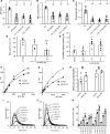
Figure 3. HETE-PL restoration of hemostasis in mice lacking FVIII and enhancement of thrombin generation in FVIII deficiency, with/without tissue factor pathway inhibitor (TFPI) inhibition.
(A and B) Summary data for thrombin generation in plasma as shown in Figure 3, E–L (n = 3, mean ± SEM). **P < 0.01, compared with control, as determined by 1-way ANOVA and post hoc Tukey tests. (C and D) 12-HETE-PE restores hemostasis in mice lacking FVIII. Male mice (11 weeks) were administered 78 ng HETE-PE in liposomes as described in Methods before a 3-mm tail cut. Bleeding time and Hb loss were determined as in Methods (n = 4–9 mice per group). Data is shown as Tukey box plots, with 1-way ANOVA with Tukey post hoc multicomparison test. *P < 0.05, ***P < 0.005, data shown as squares in box plot are outliers (values 1.5–3 times lower or higher than the interquartile range).
Figure 4
Figure 4. HETE-PLs enhance extrinsic tenase (FVIIa/TF), intrinsic tenase (FVIIIa/FIXa), and prothrombinase (FVa/FXa) activities in vitro.
(A and B) HETE-PLs enhance extrinsic tenase activity. Liposomes (4 mM) containing 50 pmol/l TF were added to FVIIa (10 nmol/l) and FX (136 nmol/l). Liposome composition for PE was 65% DSPC, 5% SAPS, 20% SAPE, with 10% SAPE (control) or 10% HETE-PE; for PC, it was 55% DSPC, 5% SAPS, 30% SAPE, 10% SAPC (control) or 10% HETE-PC. (A) Representative traces are shown from 1 of 4 experiments. (B) Summary data showing maximal thrombin generation (n = 4 separate experiments, each in triplicate, mean ± SEM). (C and D) HETE-PLs enhance intrinsic tenase activity. FVIIIa (300 pmol/l) was generated using FIIa and then inhibited by hirudin. This was added with substrate to FIXa (80 nmol/l), FX (136 nmol/l), and liposomes (without TF), and FXa generation was measured. (C) Representative traces from 1 of 3 experiments. (D) Summary data for maximal FXa generation (n = 3, mean ± SEM). (E–H) HETE-PLs enhance prothrombinase activity. Liposomes (4 mM lipid, composition as above, with no TF) were added to FVa, FXa, and FII at 26 nmol/l, 136 nmol/l, and 1.4 μM respectively. (E and F) Representative data showing time courses for control and HETE-PL liposomes (n = 3 technical replicates, mean ± SEM). (G) Fold-changes at different lipid concentrations for HETE-PLs (differences between means, shown in H). (H) Prothrombinase activity in the presence of liposomes, at different lipid concentrations (n = 3–4, mean ± SEM) **P < 0.01, *P < 0.05, as determined by 1-way ANOVA and post hoc Tukey tests.
Figure 5
Figure 5. HETE-PLs containing membranes directly bind Gla domain proteins, and specific HETE-PL positional isomers optimally support coagulation.
(A–C) Liposomes were analyzed using DLS for ζ potential, electrophoretic mobility, and polydispersity. Liposomes (65% DSPC, 5% DSPC, 10%–0% SAPE, 0%–10% 15-HETE-PE) were analyzed using a Zetasizer Nano, as described in Methods (n = 3–5 separate liposome preparations, each analyzed as 4 replicates) and compared for differences against PC liposomes, using 1-way ANOVA with multicolumn comparison using Tukey. **P < 0.01. (D) Liposomes (65% DSPC, 5% DSPC, 30%–0% SAPE, 0%–0% 15-HETE-PE) were analyzed by nanoparticle tracking analysis to determine the mean diameter (n = 4, mean ± SEM). (E) X-ray diffraction was carried out at beamline I22 (Diamond Light Source). Samples in glass capillary tubes were held at 37°C ± 0.1°C, and images were integrated as described in Methods (n = 3, mean ± SEM). (F–H) SPR analysis of protein binding to nanodiscs demonstrates increased binding in the presence of HETE-PE. Control discs were composed of 15% SAPS, 30% SAPE, and 55% DSPC. Nanodiscs containing HETE-PE were composed of 15% SAPS, 20% SAPE, 55% DSPC, and 10% HETE-PE. (F) Representative SPR-derived binding isotherms for FX binding to HETE-PE and control nanodiscs. Discs used were of the same composition as described for A. (G) Representative SPR-derived binding isotherms for prothrombin binding to HETE-PE containing and control nanodiscs. Discs used were of the same composition as described A. (H) Molecules of protein bound per leaflet of nanodiscs in the presence or absence of HETE-PE. Summary data for experiments, n = 4 (FX), 2 (FII). (I–K) Positional isomer of HETE-PL influences potency of thrombin stimulation. Thrombin generation was initiated by addition of tissue factor containing liposomes to plasma as described in Methods. (I) liposomes contained 65% DSPC, 5% SAPS, and 30% SAPE, with 10% SAPE replaced with 10% HETE-PE. (J) Liposomes contained 55% DSPC, 5% SAPS, and 30% SAPE, with 10% SAPC replaced with 10% HETE-PC. (K) Summary data for maximum thrombin generation (n = 3, mean ± SEM). ***P < 0.005, **P < 0.01, *P < 0.05, as determined by 1-way ANOVA and post hoc Tukey tests.
Figure 6
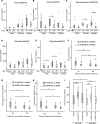
Figure 6. Altered generation and action of HETE-PLs in a clinical setting of hemostatic failure associated with cardiopulmonary bypass surgery.
(A–E) Platelets after CPB generate less HETE-PE and externalize less HETE-PE and PS than those isolated before CPB. Platelets isolated from peripheral blood of patients, before (shaded) and after (unshaded) CPB were measured for HETE-PE, HETE-PC, and externalized PE, PS, and HETE-PE basally and following thrombin or collagen activation, as described in Methods (n = 12). (F–H) Stimulation of thrombin generation in preoperative plasma by 12-HETE-PL was significantly reduced in patients who received hemostatic treatment after CPB. Plasma from patients before CPB was added to 50 pmol/l TF liposomes with/without 10% HETE-PL, and thrombin was measured as described in Methods. ETP (F), peak thrombin (G), and velocity index (H) were determined and compared with responses to liposomes that contained native PL, to give fold changes for each. (I) Reduced thrombin generation seen in post-CPB plasma was restored using HETE-PL. Thrombin generation was compared in plasma before or after CPB using liposomes containing HETE-PL, as indicated (10%). For A–I, horizontal black lines represent median, boxes represent the interquartile range, bars represent values falling within 1.5 times the interquartile range, and asterisks/circles represent outliers (values 1.5–3 times lower or higher than the interquartile range). n = 87 for panels F–I. Data were analyzed using Wilcoxon rank test for paired variables, Mann Whitney U test for unpaired variables and Friedman’s 2-way analysis of variance with post hoc testing to compare repeated measures of related variables, **P < 0.01, *P < 0.05.

Similar articles

Cited by

References

    1. Mann KG. Biochemistry and physiology of blood coagulation. Thromb Haemost. 1999;82(2):165–174. - PubMed
    1. Morrissey JH, Tajkhorshid E, Rienstra CM. Nanoscale studies of protein-membrane interactions in blood clotting. J Thromb Haemost. 2011;9 Suppl 1:162–167. - PMC - PubMed
    1. Zwaal RF, Schroit AJ. Pathophysiologic implications of membrane phospholipid asymmetry in blood cells. Blood. 1997;89(4):1121–1132. - PubMed
    1. Taylor FB, et al. Lethal E. coli septic shock is prevented by blocking tissue factor with monoclonal antibody. Circ Shock. 1991;33(3):127–134. - PubMed
    1. Levi M, et al. Inhibition of endotoxin-induced activation of coagulation and fibrinolysis by pentoxifylline or by a monoclonal anti-tissue factor antibody in chimpanzees. J Clin Invest. 1994;93(1):114–120. doi: 10.1172/JCI116934. - DOI - PMC - PubMed

Publication types

MeSH terms