. 2018 Mar 22;3(6):e99249.
doi: 10.1172/jci.insight.99249.

TNF and granulocyte macrophage-colony stimulating factor interdependence mediates inflammation via CCL17

Affiliations

TNF and granulocyte macrophage-colony stimulating factor interdependence mediates inflammation via CCL17

Andrew D Cook et al. JCI Insight. .

Abstract

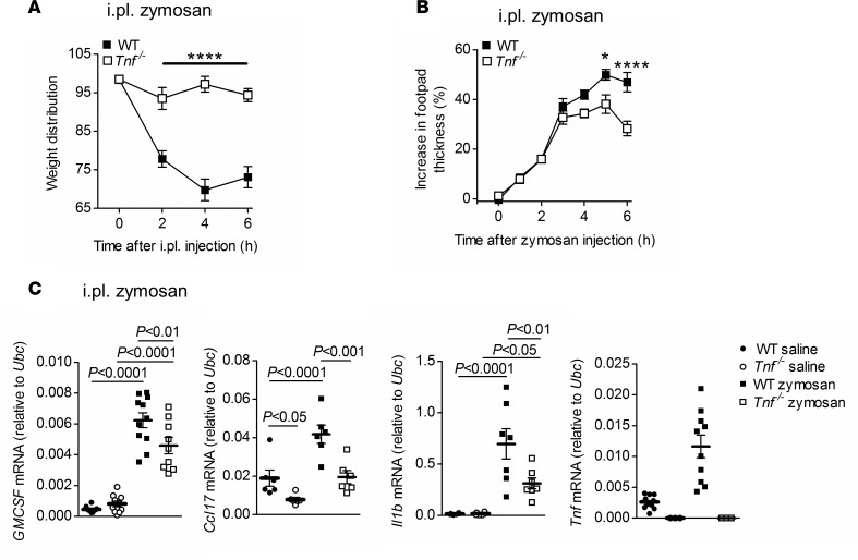
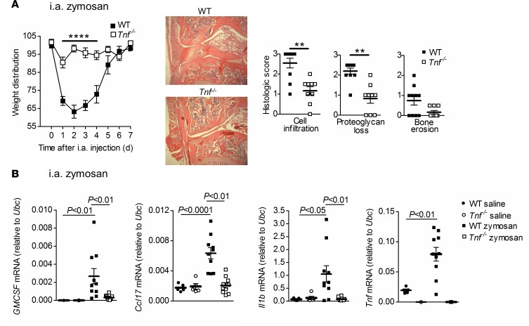
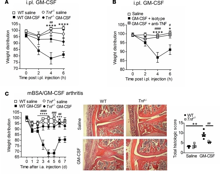
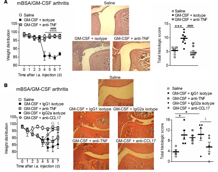
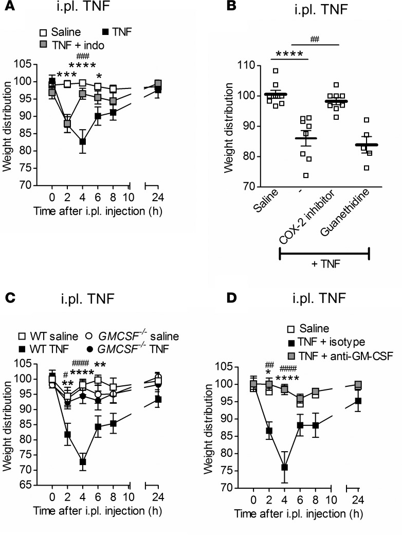
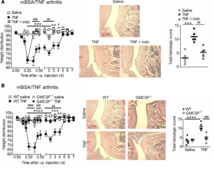
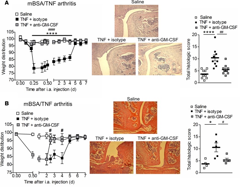
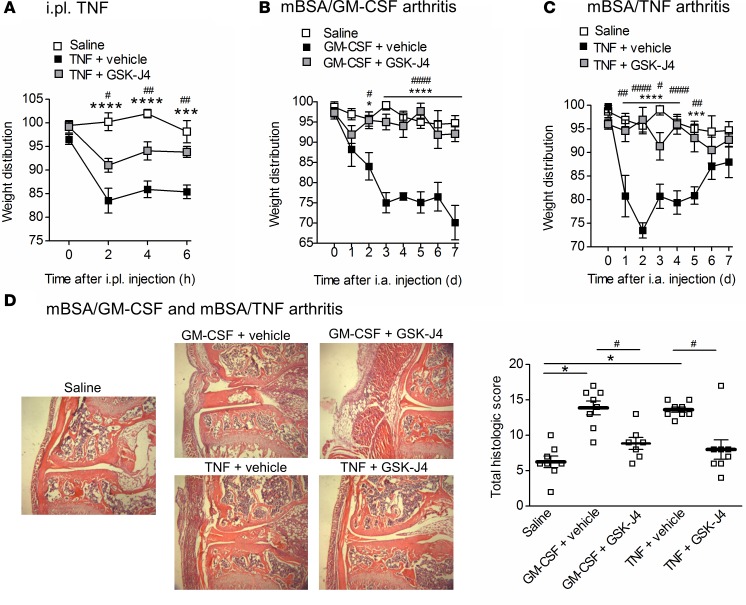
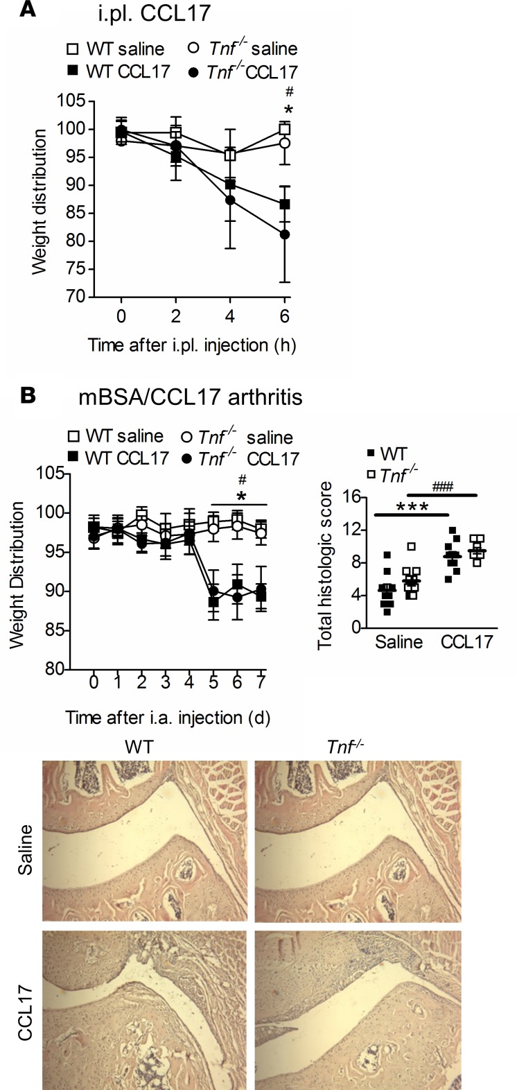
TNF and granulocyte macrophage-colony stimulating factor (GM-CSF) have proinflammatory activity and both contribute, for example, to rheumatoid arthritis pathogenesis. We previously identified a new GM-CSF→JMJD3 demethylase→interferon regulatory factor 4 (IRF4)→CCL17 pathway that is active in monocytes/macrophages in vitro and important for inflammatory pain, as well as for arthritic pain and disease. Here we provide evidence for a nexus between TNF and this pathway, and for TNF and GM-CSF interdependency. We report that the initiation of zymosan-induced inflammatory pain and zymosan-induced arthritic pain and disease are TNF dependent. Once arthritic pain and disease are established, blockade of GM-CSF or CCL17, but not of TNF, is still able to ameliorate them. TNF is required for GM-CSF-driven inflammatory pain and for initiation of GM-CSF-driven arthritic pain and disease, but not once they are established. TNF-driven inflammatory pain and TNF-driven arthritic pain and disease are dependent on GM-CSF and mechanistically require the same downstream pathway involving GM-CSF→CCL17 formation via JMJD3-regulated IRF4 production, indicating that GM-CSF and CCL17 can mediate some of the proinflammatory and algesic actions of TNF. Given we found that TNF appears important only early in arthritic pain and disease progression, targeting a downstream mediator, such as CCL17, which appears to act throughout the course of disease, could be effective at ameliorating chronic inflammatory conditions where TNF is implicated.

Keywords: Arthritis; Cytokines; Inflammation; Macrophages.


Conflict of interest statement

Conflict of interest: JES is an employee of GSK Research and Development Limited.

Figures

Figure 1
Figure 1. TNF is required for zymosan-induced inflammatory pain.
(AC) WT and Tnf–/– mice received an intraplantar (i.pl.) injection of zymosan, and (A) pain (incapacitance meter [ratio of weight bearing on injected relative to noninjected hindlimb]; a value < 100 indicates pain), (B) swelling, and (C) footpad mRNA expression (6 hours zymosan or saline) were measured (n = 6–10 mice/group). Results are shown as mean ± SEM. P values were obtained using a 2-way ANOVA. *P < 0.05, ****P < 0.0001, WT versus Tnf–/– mice.
Figure 2
Figure 2. TNF is required for zymosan-induced arthritic pain and optimal disease.
(A and B) WT and Tnf–/– mice received an intra-articular (i.a.) injection of zymosan; (A) pain and arthritis (histology, day 7) and (B) joint mRNA expression (day 7 zymosan or saline) were measured (n = 5–10 mice/group). For histology images, original magnification ×60. Results are shown as mean ± SEM. P values were obtained using a 2-way ANOVA test for pain (weight distribution) readings (A) and gene expression (B), and Mann-Whitney U test for histology (A). **P < 0.01, ****P < 0.0001, WT versus Tnf–/– mice.
Figure 3
Figure 3. TNF is not required for the maintenance of zymosan-induced arthritic pain and disease.
(A and B) WT mice received an intra-articular (i.a.) injection of zymosan and anti-TNF mAb, anti–granulocyte macrophage-colony stimulating factor (anti–GM-CSF) mAb, IgG1 or IgG2a isotype control mAbs (150 μg i.p.), either (A) prophylactically (on days –2 and 0) (n = 4–5 mice/group) or (B) therapeutically (day 1) (n = 5–10 mice/group), and pain and arthritis (histology, day 7) were measured. Mice were also treated (B) therapeutically with anti-CCL17 mAb (150 μg i.p.) (day 1) (n = 5 mice/group). For histology images, original magnification ×60. Results are shown as mean ± SEM. P values were obtained using a 2-way ANOVA test for pain (weight distribution) readings, and a 1-way ANOVA test for histology. *P < 0.05, ***P < 0.001, ****P < 0.0001, anti-TNF versus IgG1 isotype; #P < 0.05, ##P < 0.01, ###P < 0.001, ####P < 0.0001, anti–GM-CSF versus IgG2a isotype; ζP < 0.05, ζζP < 0.01, anti-CCL17 versus IgG2a isotype.
Figure 4
Figure 4. GM-CSF–driven inflammatory pain, as well as GM-CSF–driven arthritic pain and disease, are TNF dependent.
(A and B) Intraplantar (i.pl.) injection of granulocyte macrophage-colony stimulating factor (GM-CSF) (20 ng) or saline in (A) WT and Tnf–/– mice (n = 4–6 mice/group) and in (B) WT mice treated with anti-TNF or isotype control (2 μg/paw i.pl. at t = 0) (n = 12 mice/group). Pain was measured. (C) Methylated BSA (mBSA)/GM-CSF arthritis (intra-articular [i.a.] mBSA [day 0] and GM-CSF or saline s.c. [days 0–2]) was induced in WT and Tnf–/– mice. Pain and arthritis (histology, day 7) were measured (n = 4–6 mice/group). Original magnification, ×60. Results are shown as mean ± SEM. P values were obtained using a 2-way ANOVA. *P < 0.05, **P < 0.01, ***P < 0.001, ****P < 0.0001, WT saline versus WT GM-CSF; #P < 0.05, ##P < 0.01, ###P < 0.001, ####P < 0.0001, WT GM-CSF versus Tnf–/– GM-CSF.
Figure 5
Figure 5. Prophylactic, but not therapeutic, treatment with anti-TNF mAb ameliorates GM-CSF–driven arthritic pain and disease.
(A and B) Methylated BSA (mBSA)/granulocyte macrophage-colony stimulating factor (GM-CSF) arthritis (intra-articular [i.a.] mBSA [day 0] and GM-CSF or saline s.c. [days 0–2]) was induced in WT mice treated with anti-TNF mAb or IgG1 isotype control (150 μg i.p.) (A) prophylactically (on days –2 and 0) (n = 8–12 mice/group) and (B) therapeutically (day 4) (n = 5 mice/group). Mice were also treated (B) therapeutically with anti-CCL17 mAb or IgG2a isotype control (150 μg i.p.) (day 4) (n = 4–5 mice/group). Pain (incapacitance meter) and arthritis (histology) were measured. Original magnification, ×125. Results are shown as mean ± SEM. P values were obtained using a 2-way ANOVA test for pain (weight distribution) readings and a Mann-Whitney U test or 1-way ANOVA for histology quantification. *P < 0.05, ***P < 0.001, ****P < 0.0001, saline versus GM-CSF + isotype; ####P < 0.0001, GM-CSF + isotype versus GM-CSF + anti-TNF; ζP < 0.05, ζζP < 0.01, GM-CSF + isotype versus GM-CSF + anti-CCL17.
Figure 6
Figure 6. GM-CSF is required for TNF-driven inflammatory pain.
(AD) Intraplantar (i.pl.) injection of TNF (20 ng) or saline in (A) WT mice treated with/without indomethacin (12.5 μg/paw i.pl. at 2 hours) (n = 10 mice/group), (B) WT mice treated with/without the COX-2 inhibitor, SC58125 (5 mg/kg i.p. at t = –30 minutes), or guanethidine sulfate (25 mg/kg s.c. at –60 minutes) for 4 hours (n = 5–8 mice/group), (C) WT and GMCSF–/– mice (n = 5–10 mice/group), and (D) WT mice treated with anti–GM-CSF mAb or isotype control (2 μg/paw i.pl. at t = 0) (n = 5 mice/group). Pain (incapacitance meter) was measured. Results are shown as mean ± SEM. P values were obtained using a 1-way (B) or 2-way (A, C, and D) ANOVA test. *P < 0.05, **P < 0.01, ***P < 0.001, ****P < 0.0001, WT saline versus WT TNF or TNF + isotype; #P < 0.05, ##P < 0.01, ###P < 0.001, ####P < 0.0001, TNF versus TNF + indo or TNF + COX-2 inhibitor; WT TNF versus GMCSF–/– TNF; TNF + isotype vs. TNF + anti–GM-CSF. GM-CSF, granulocyte macrophage-colony stimulating factor.
Figure 7
Figure 7. GM-CSF is required for TNF-driven arthritic pain and disease.
(A and B) Methylated BSA (mBSA)/TNF arthritis (intra-articular [i.a.]mBSA [day 0] and TNF or saline s.c. [days 0–2]) was induced in (A) WT mice treated with/without indomethacin (1 mg/kg, i.p. from 6 hours) (n = 7–10 mice/group), and (B) GMCSF–/– mice (n = 8–10 mice/group). Pain and arthritis (histology) were measured. Original magnification, ×125. Results are shown as mean ± SEM. P values were obtained using a 2-way ANOVA test for pain (weight distribution) readings, a 1-way (A) or 2-way (B) ANOVA test for histology quantification. *P < 0.05, **P < 0.01, ***P < 0.001, ****P < 0.0001, WT saline versus WT TNF; #P < 0.05, ##P < 0.01, ###P < 0.001, ####P < 0.0001, TNF versus TNF + indo, WT TNF versus GMCSF–/– TNF. GM-CSF, granulocyte macrophage-colony stimulating factor.
Figure 8
Figure 8. Prophylactic and therapeutic treatment with anti–GM-CSF mAb ameliorates TNF-driven arthritic pain and disease.
(A and B) Methylated BSA (mBSA)/TNF arthritis (intra-articular [i.a.] mBSA [day 0] and TNF or saline s.c. [days 0–2]) was induced in (A) WT mice treated prophylactically with anti–granulocyte macrophage-colony stimulating factor (anti–GM-CSF) mAb or isotype control (150 μg i.p. on days –2 and 0 i.p.) (n = 9–10 mice/group) and (B) WT mice treated therapeutically with anti–GM-CSF mAb or isotype control (150 μg i.p. on day 2 i.p.) (n = 4–5 mice/group). Pain and arthritis (histology) were measured. Original magnification, ×125. Results are shown as mean ± SEM. P values were obtained using a 2-way ANOVA test for pain (weight distribution) readings, and a 1-way ANOVA test for histology quantification. *P < 0.05, ****P < 0.0001, saline versus TNF + isotype; #P < 0.05, ##P < 0.01, ####P < 0.0001, TNF + isotype versus TNF + anti–GM-CSF.
Figure 9
Figure 9. CCL17 is required for TNF-driven arthritic pain and disease.
(A and B) Methylated BSA (mBSA)/TNF arthritis (intra-articular [i.a.] mBSA [day 0] and TNF or saline s.c. [days 0–2]) was induced in (A) WT and Ccl17E/E mice (n = 5–6 mice/group) and (B) WT mice treated therapeutically with anti-CCL17 mAb or isotype control (150 μg i.p. on day 2 i.p.) (n = 4–5 mice/group). Pain and arthritis (histology) were measured. Original magnification, ×125. Results are shown as mean ± SEM. P values were obtained using a 2-way ANOVA test for pain and a 1-way (B) or 2-way (A) ANOVA test for histology quantification. *P < 0.05, **P < 0.01, ***P < 0.001, WT saline versus WT TNF; #P < 0.05, ###P < 0.001, WT TNF versus Ccl17E/E TNF; TNF + isotype versus TNF + anti-CCL17.
Figure 10
Figure 10. Inhibition of JMJD3 demethylase ameliorates TNF-driven inflammatory pain, and GM-CSF– and TNF-driven arthritic pain and disease.
(A) Intraplantar (i.pl.) injection of TNF (20 ng) or saline in WT mice treated with vehicle or GSK-J4 (25 mg/kg i.p. at t = –30 minutes). Pain was measured (n = 4–7 mice/group). (BD) Methylated BSA (mBSA)/granulocyte macrophage-colony stimulating factor (GM-CSF) arthritis and mBSA/TNF arthritis were induced in WT mice treated with vehicle or GSK-J4 (0.5 mg/kg i.p. daily from day –2 to day 2). (B and C) Pain and (D) arthritis (histology) were measured (n = 7–8 mice/group). Original magnification, ×60. Results are shown as mean ± SEM. P values were obtained using a 2-way ANOVA test for pain and a 1-way (D) ANOVA test for histology quantification. *P < 0.05, ***P < 0.001, ****P < 0.0001, saline versus TNF or GM-CSF + vehicle; #P < 0.05, ##P < 0.01, ####P < 0.0001, vehicle versus GSK-J4.
Figure 11
Figure 11. TNF is not required for CCL17-driven inflammatory pain and CCL17-driven arthritic pain and disease.
(A) Pain (incapacitance meter) was measured in WT and Tnf–/– mice following intraplantar (i.pl.) injection of CCL17 (50 ng) or saline (n = 5 mice/group). (B) Methylated BSA (mBSA)/CCL17 arthritis (intra-articular [i.a.] mBSA [day 0] and CCL17 or saline [days 0–2]) was induced in WT and Tnf–/– mice and pain (incapacitance meter) and arthritis (histology) were measured (n = 6–10 mice/group). Original magnification, ×125. Results are shown as mean ± SEM. P values were obtained using 2-way ANOVA test. *P < 0.05, ***P < 0.001, WT saline versus WT CCL17; #P < 0.05, ###P < 0.001, Tnf–/– saline versus Tnf–/– CCL17.

Similar articles

Cited by

References

    1. Noack M, Miossec P. Selected cytokine pathways in rheumatoid arthritis. Semin Immunopathol. 2017;39(4):365–383. - PubMed
    1. Hamilton JA. Colony-stimulating factors in inflammation and autoimmunity. Nat Rev Immunol. 2008;8(7):533–544. doi: 10.1038/nri2356. - DOI - PubMed
    1. Hamilton JA. GM-CSF as a target in inflammatory/autoimmune disease: current evidence and future therapeutic potential. Expert Rev Clin Immunol. 2015;11(4):457–465. doi: 10.1586/1744666X.2015.1024110. - DOI - PubMed
    1. Hamilton JA, Cook AD, Tak PP. Anti-colony-stimulating factor therapies for inflammatory and autoimmune diseases. Nat Rev Drug Discov. 2016;16(1):53–70. - PubMed
    1. Burmester GR, et al. A randomised phase IIb study of mavrilimumab, a novel GM-CSF receptor alpha monoclonal antibody, in the treatment of rheumatoid arthritis. Ann Rheum Dis. 2017;76(6):1020–1030. doi: 10.1136/annrheumdis-2016-210624. - DOI - PubMed

Publication types

MeSH terms