Review
. 2016 Sep 6;4(3):30.
doi: 10.3390/dj4030030.

Negative Pressure Wound Therapy in Maxillofacial Applications

Affiliations
Review

Negative Pressure Wound Therapy in Maxillofacial Applications

Adam J Mellott et al. Dent J (Basel). .

Abstract

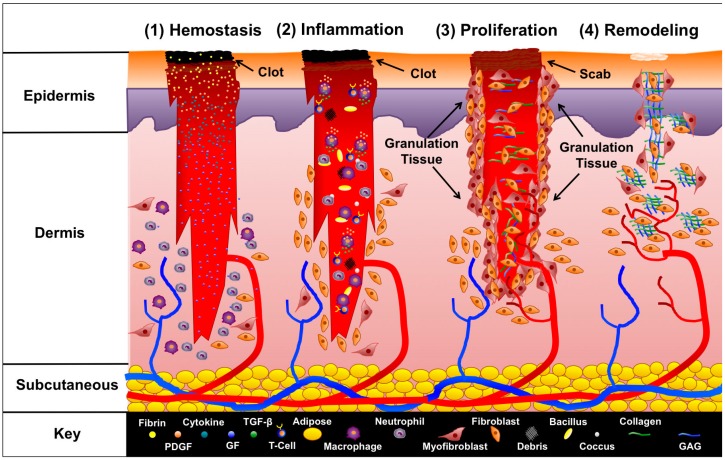
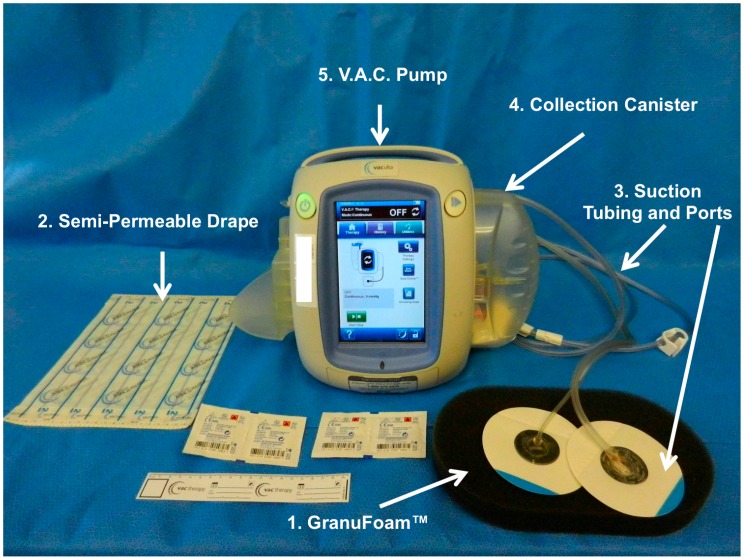
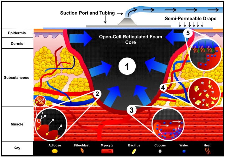
Negative pressure wound therapy has greatly advanced the field of wound healing for nearly two decades, by providing a robust surgical adjunct technique for accelerating wound closure in acute and chronic wounds. However, the application of negative pressure wound therapy in maxillofacial applications has been relatively under utilized as a result of the physical articulations and contours of the head and neck that make it challenging to obtain an airtight seal for different negative pressure wound therapy systems. Adapting negative pressure wound therapies for maxillofacial applications could yield significant enhancement of wound closure in maxillofacial applications. The current review summarizes the basic science underlying negative pressure wound therapy, as well as specific maxillofacial procedures that could benefit from negative pressure wound therapy.

Keywords: craniofacial; maxillofacial; negative pressure wound therapy; vacuum assisted closure; wound healing.


Conflict of interest statement

D.S.Z. declares that he has sold patents to the V.A.C. to KCI/Acelity and continues to receive royalties on Prevena (KCI/Acelity). A.J.M. and B.T.A. declare no conflicts of interest.

Figures

Figure 1
Figure 1
Phases of wound healing. The four phases of wound healing are illustrated. Immediately after a wound forms, hemostasis begins. Platelets form a clot, and are bound together by fibrin. Several cytokines are released, which recruit neutrophils and other leukocytes to the site of injury to start the inflammation phase. Leukocytes begin clearing the wound of bacteria, debris, and other foreign contaminants. T-cells infiltrate the wound and recruit macrophages, which release PDGF and TGF-β to signal fibroblasts and myofibroblasts to start the proliferation phase. Granulation tissue forms, and fibroblasts begin developing new extracellular fibers by producing collagen and GAGs. In addition angiogenesis begins, and new blood is supplied to the site of injury. After proliferation, the final phase, remodeling, occurs in which extracellular fibers align, the wound contracts, and fibroblasts release enzymes to remove damaged extraneous extracellular matrix. Epithelial cells, despite their defining role in coverage, healing, and controlling “crosstalk” with fibroblasts, have been deliberately left out of this schematic representation for the sake of clarity. (PDGF = Platelet-Derived Growth Factor, GF = Growth Factor, GAG = Glycosaminoglycan).
Figure 2
Figure 2
Vacuum Assisted Closure (V.A.C.) system. KCI Veraflow™ is the flagship model, and is designed to provide instillation therapy in addition to standard V.A.C. therapy. The (1) black semi-occlusive dressing (GranuFoam™) along with the (2) semi-permeable drape used to isolate the wound and prevent escape of evaporative moisture. In addition, the (3) suction tubing and suction ports are connected to the (4) collection canister through which (5) the vacuum pump exerts suction force.
Figure 3
Figure 3
V.A.C. mechanisms of action. An open-cell reticulated foam is placed within the wound and covered by a semi-permeable adhesive drape. The drape is adhesively secured around and over the wound to create an airtight seal. A small hole is made within the center of the drape, and the suction port and tubing connected to the collection canister are attached. Engaging the vacuum pump evacuates the air from the foam and enables (1) Macrodeformation of the foam via shrinkage, which pulls the wound edges together. At the interface between the foam and wound bed, (2) microstrain occurs in which cells are pulled into the pores of the foam while an equal and opposing force acting on the struts of the foam pushes cells away. The microstrain on the cells initiates mechanotransduction, which can stimulate cell proliferation as illustrated within the sub-inset of inset 2. Additionally, engagement of the vacuum facilitates the (3) movement of fluid out of interstitial spaces, thereby reducing edema and increasing blood flow as illustrated in inset 3. The V.A.C. system possibly (4) reduces bacterial burden; however, the mechanism by which bacteria are reduced is not fully understood. The destruction of bacteria is illustrated in inset 4. The V.A.C. system contributes to (5) wound stabilization through secondary events. Inset 5 illustrates the movement of warm air down through the semi-permeable drape into the wound space, while isolating the wound from foreign contaminants. Furthermore, the semi-permeable drape prevents evaporative water loss, which aids in keeping the wound moist to enable cell migration and nutrient transport.

Similar articles

Cited by

References

    1. Banwell P., Withey S., Holten I. The use of negative pressure to promote healing. Br. J. Plast. Surg. 1998;51:79. doi: 10.1016/S0007-1226(98)80142-2. - DOI - PubMed
    1. Orgill D.P., Bayer L.R. Update on negative-pressure wound therapy. Plast. Reconstr. Surg. 2011;127(Suppl. 1):105S–115S. doi: 10.1097/PRS.0b013e318200a427. - DOI - PubMed
    1. Asher S.A., White H.N., Golden J.B., Magnuson J.S., Carroll W.R., Rosenthal E.L. Negative pressure wound therapy in head and neck surgery. JAMA Facial Plast. Surg. 2014;16:120–126. doi: 10.1001/jamafacial.2013.2163. - DOI - PubMed
    1. Armstrong D.G., Andros G. Use of negative pressure wound therapy to help facilitate limb preservation. Int. Wound J. 2012;9(Suppl. 1):1–7. doi: 10.1111/j.1742-481X.2012.01015.x. - DOI - PMC - PubMed
    1. Dumville J.C., Land L., Evans D., Peinemann F. Negative pressure wound therapy for treating leg ulcers. Cochrane Database Syst. Rev. 2015;7:CD011354. - PMC - PubMed

LinkOut - more resources