. 2018 Mar 21;8(3):35.
doi: 10.1038/s41408-018-0062-y.

A multiple myeloma-specific capture sequencing platform discovers novel translocations and frequent, risk-associated point mutations in IGLL5

Affiliations

A multiple myeloma-specific capture sequencing platform discovers novel translocations and frequent, risk-associated point mutations in IGLL5

Brian S White et al. Blood Cancer J. .

Abstract

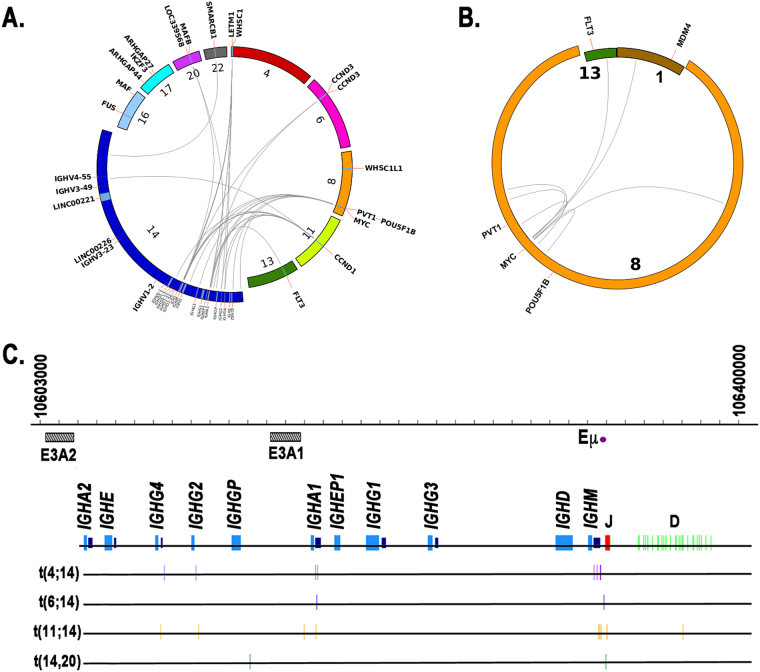
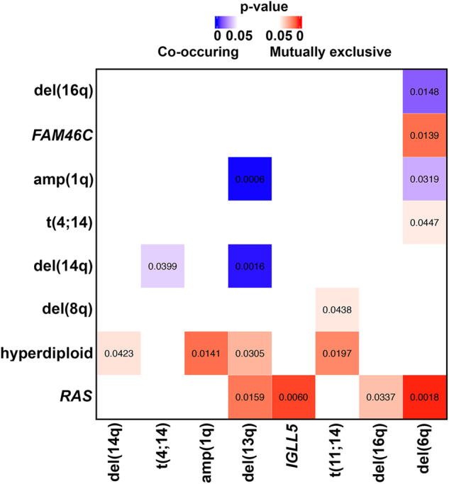
Multiple myeloma (MM) is a disease of copy number variants (CNVs), chromosomal translocations, and single-nucleotide variants (SNVs). To enable integrative studies across these diverse mutation types, we developed a capture-based sequencing platform to detect their occurrence in 465 genes altered in MM and used it to sequence 95 primary tumor-normal pairs to a mean depth of 104×. We detected cases of hyperdiploidy (23%), deletions of 1p (8%), 6q (21%), 8p (17%), 14q (16%), 16q (22%), and 17p (4%), and amplification of 1q (19%). We also detected IGH and MYC translocations near expected frequencies and non-silent SNVs in NRAS (24%), KRAS (21%), FAM46C (17%), TP53 (9%), DIS3 (9%), and BRAF (3%). We discovered frequent mutations in IGLL5 (18%) that were mutually exclusive of RAS mutations and associated with increased risk of disease progression (p = 0.03), suggesting that IGLL5 may be a stratifying biomarker. We identified novel IGLL5/IGH translocations in two samples. We subjected 15 of the pairs to ultra-deep sequencing (1259×) and found that although depth correlated with number of mutations detected (p = 0.001), depth past ~300× added little. The platform provides cost-effective genomic analysis for research and may be useful in individualizing treatment decisions in clinical settings.


Conflict of interest statement

The authors declare that they have no conflict of interest.

Figures

Fig. 1
Fig. 1. Targeted sequencing identifies chromosome-level, arm-level, and focal CNVs.
a Hyperdiploid and (b; arrow) focal copy number events detected by CopyCat2 (blue; p < 0.05) from log2 ratios of tumor to paired normal sequencing depth (y-axis) across chromosomes (x-axis). (Clonal) single-copy gains occur at a log2 ratio of log2(3/2)  =0.58, whereas (clonal) heterozygous/single-copy losses occur at a log2 ratio of log2(1/2) = −1
Fig. 2
Fig. 2. Targeted sequencing detects IGH and MYC translocations.
Circos plots of (a) IGH and (b) MYC translocations. Chromosomes involved in translocations are magnified to highlight regions and genes near breakpoints. c Breakpoints (vertical lines) of canonical IGH translocations within IGH locus. E3A2 and E3A1: 3’ enhancer elements downstream of IGHA2 and IGHA1 genes, respectively. Eμ: μ enhancer. Purple boxes: switch regions. Figure is to scale
Fig. 3
Fig. 3. IGLL5 is translocated and DERL3 is overexpressed in multiple myeloma.
a Schematic of validated t(14;22) translocation. (Left) WT chromosomes 14 and 22 with horizontal lines indicating location of breakpoints within the IGH and IGLL5 loci, respectively. (Right) Two derivative (der) chromosomes, each retaining a portion of its respective IGH or IGLL5 gene. Cancer-associated genes within 1 Mb of breakpoint on der(14) are shown. b PCR validation of t(14,22) translocation. Oligos specific to each breakpoint used in PCR reactions (top). Oligos specific to the small regions deleted on der(14) and der(22) were designed to detect non-translocated allele (bottom). T tumor, N germline (peripheral blood) control. cDERL3 expression across 84 MM patients. Red circle indicates sample in which putative (non-validated) t(14;22) translocation was detected. FPKM Fragments Per Kilobase of transcript per Million mapped reads
Fig. 4
Fig. 4. Greater sequencing depth yields few additional variants.
VAF of variants discovered during initial targeted sequencing (x-axis) and/or with subsequent deeper sequencing (y-axis)
Fig. 5
Fig. 5. Targeted sequencing identifies CNVs, SNVs, and translocations.
a Mutations per Mb, b SNVs, and c CNVs and translocations detected across 95 samples (columns). MYC-ITX: intra-chromosomal MYC translocations; MYC-CTX: inter-chromosomal MYC translocations; Non MYC-IGH CTX: inter-chromosomal IGH translocations, excluding those involving MYC
Fig. 6
Fig. 6. Targeted sequencing identifies co-occurrence and mutual exclusivity across mutation types.
Co-occurring (blue) and mutually exclusive (red) mutations (p < 0.05). Numbers indicate p-values
Fig. 7
Fig. 7. IGLL5 mutations are associated with increased risk of disease progression.
Kaplan-Meier curves of IGLL5 mutant samples (with non-synonymous SNVs and/or indels) versus IGLL5 WT samples

References

    1. Manier, S. et al. Genomic complexity of multiple myeloma and its clinical implications. Nat. Rev. Clin. Oncol. 14, 100–103 (2016). - PubMed
    1. Morgan GJ, Walker BA, Davies FE. The genetic architecture of multiple myeloma. Nat. Rev. Cancer. 2012;12:335–348. doi: 10.1038/nrc3257. - DOI - PubMed
    1. Greipp PR, et al. International staging system for multiple myeloma. J. Clin. Oncol. 2005;23:3412–3420. doi: 10.1200/JCO.2005.04.242. - DOI - PubMed
    1. Walker BA, et al. Mutational spectrum, copy number changes, and outcome: results of a sequencing study of patients with newly diagnosed myeloma. J. Clin. Oncol. 2015;33:3911–3920. doi: 10.1200/JCO.2014.59.1503. - DOI - PMC - PubMed
    1. Chretien ML, et al. Understanding the role of hyperdiploidy in myeloma prognosis: which trisomies really matter? Blood. 2015;126:2713–2719. doi: 10.1182/blood-2015-06-650242. - DOI - PMC - PubMed

Publication types