. 2018 Mar 21;8(1):4975.
doi: 10.1038/s41598-018-23176-x.

The evolutionarily conserved genes: Tex37, Ccdc73, Prss55 and Nxt2 are dispensable for fertility in mice

Affiliations

The evolutionarily conserved genes: Tex37, Ccdc73, Prss55 and Nxt2 are dispensable for fertility in mice

Manan Khan et al. Sci Rep. .

Abstract

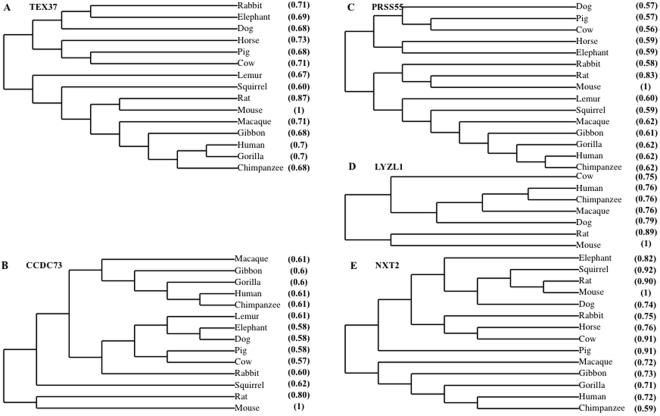
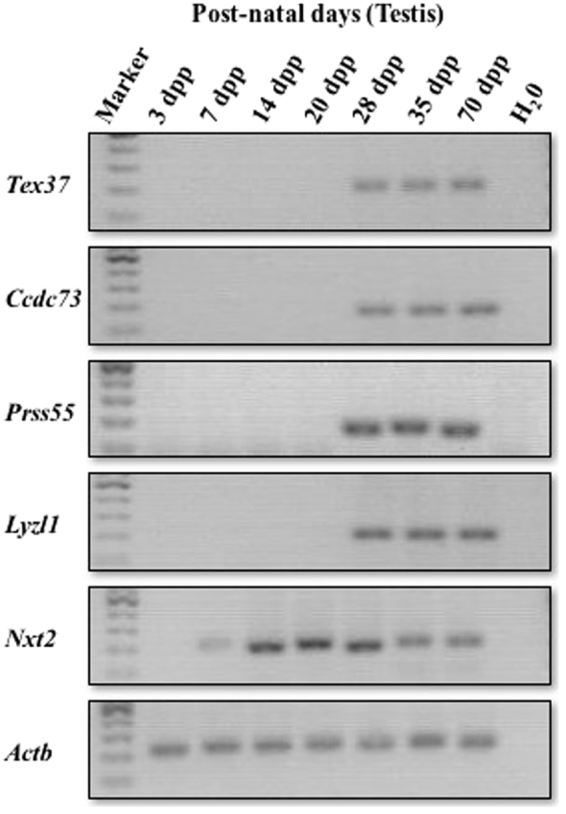
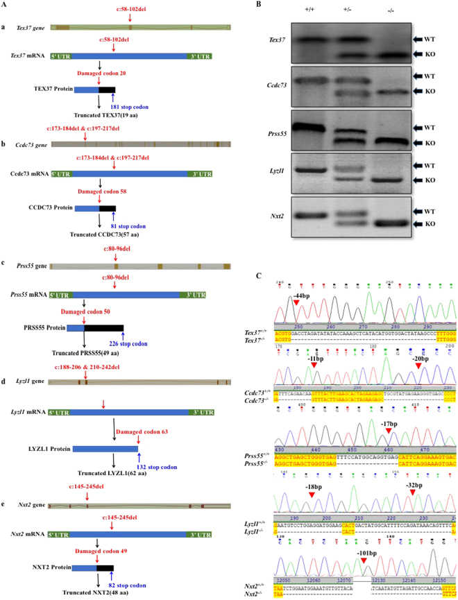
There are more than 2300 genes that are predominantly expressed in mouse testes. The role of hundreds of these genes has been studied in mouse spermatogenesis but still there are many genes whose function is unknown. Gene knockout (KO) strategy in mice is widely used for in vivo study of gene function. The present study was designed to explore the function of the four genes: Tex37, Ccdc73, Prss55 and Nxt2, which were evolutionarily conserved in eutherians. We found that these genes had a testis-enriched expression pattern in mice except Nxt2. We knocked out these genes by CRISPR/Cas9 individually and found that all the KO mice had normal fertility with no detectable difference in testis/body weight ratios, epididymal sperm counts, as well as testicular and epididymal histology from wild type mice. Although these genes are evolutionarily conserved in eutherians including human and mouse, they are not individually essential for spermatogenesis, testis development and male fertility in mice in laboratory conditions. Our report of these fertile KO data could avoid the repetition and duplication of efforts which will help in prioritizing efforts to focus on genes that are indispensable for male reproduction.


Conflict of interest statement

The authors declare no competing interests.

Figures

Figure 1
Figure 1
Conservation of selected genes in eutherians. Multiple proteins sequence alignments were performed by T-coffee. Phylogenetic trees were constructed using online database TreeDyn from multiple protein sequence alignment. Phylogeny of (A) Tex37, (B) Ccdc73, (C) Prss55, (D) Lyzl1, and (E) Nxt2. Parentheses show percent identity to reference sequence (mouse:1).
Figure 2
Figure 2
mRNA expression of the selected genes. Postnatal temporal expression of genes in testes of 3, 7, 14, 20, 28, 35 and 70-dpp-old mice was analyzed by RT-PCR. Actb was used as positive control. H20 was used as negative control. Cropped gels are shown here. Full-length gels are provided for review in the supplementary Figure S2a.
Figure 3
Figure 3
Knockout strategy and genotyping of mutant mice. (A) Schematic strategies for the generation of KO mice using CRISPR/Cas9. 44 bp of Tex37 (a), 11 bp and 20 bp of Ccdc73 (b) and 17 bp of Prss55 (c) were deleted from Exon 2. While, 18 bp and 32 bp of Lyzl1 (d) and 101 bp of Nxt2 (e) were deleted from exon 3. (B) Genotype of each KO mouse was confirmed with PCR. (C) Representative Sanger sequence image for the verification of each KO mouse. Red arrow heads above the chromatograms and dashes in aligned cds sequences show the deletions.
Figure 4
Figure 4
Fertility and spermatogenesis of the KO mice. (A) Representative images of testes from 70-dpp-old WT and KO mice. Scale bars, 2 mm. (B) Testis/body weight ratio of 70-dpp-old WT and KO mice. Error bars represent SD. n, the number of animals. NS, no significant difference, Student’s t-test was performed between WT and each KO mouse group for testis/body weight ratio. (C) Sperm count of 70-dpp-old WT and KO mice. Error bars represent SD. n, the number of animals. NS, no significant difference, Student’s t-test was performed between WT and each KO mouse group for sperm count. (D) H&E staining of testes and epididymides caput and cauda from 70-dpp-old WT and KO mice. Scale bars, 100μm. The data shown is representative images from at least three mice.

References

    1. Chocu S, Calvel P, Rolland AD, Pineau C. Spermatogenesis in mammals: proteomic insights. Systems biology in reproductive medicine. 2012;58:179–190. doi: 10.3109/19396368.2012.691943. - DOI - PubMed
    1. Schultz N, Hamra FK, Garbers DL. A multitude of genes expressed solely in meiotic or postmeiotic spermatogenic cells offers a myriad of contraceptive targets. Proceedings of the National Academy of Sciences. 2003;100:12201–12206. doi: 10.1073/pnas.1635054100. - DOI - PMC - PubMed
    1. Matzuk, M. M. & Lamb, D. J. Genetic dissection of mammalian fertility pathways. Translocations 45, 46XY (2002). - PubMed
    1. O’Bryan, M. K. & de Kretser, D. Mouse models for genes involved in impaired spermatogenesis. Int J Androl 29, 76–89; discussion 105–108, 10.1111/j.1365-2605.2005.00614.x (2006). - PubMed
    1. Jiang X-H, et al. Blood-testis barrier and spermatogenesis: lessons from genetically-modified mice. Asian journal of andrology. 2014;16:572. doi: 10.4103/1008-682X.125401. - DOI - PMC - PubMed

Publication types