. 2018 Mar 21;8(1):4967.
doi: 10.1038/s41598-018-23311-8.

Applicability of Automated Cell Counter with a Chlorophyll Detector in Routine Management of Microalgae

Affiliations

Applicability of Automated Cell Counter with a Chlorophyll Detector in Routine Management of Microalgae

Toshiyuki Takahashi. Sci Rep. .

Abstract

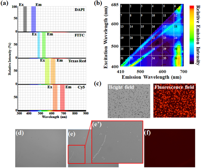
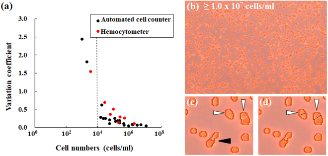
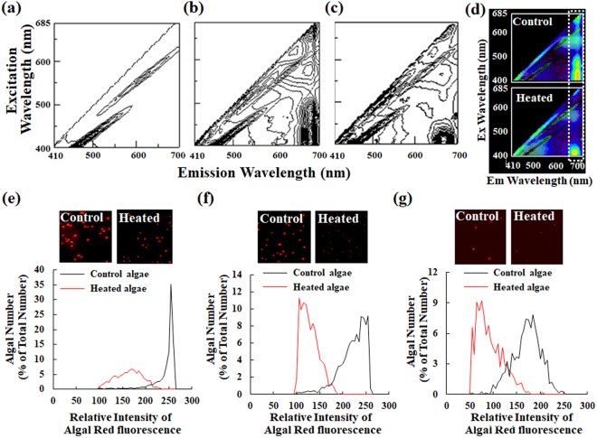
Microalgae have attracted attention for several industrial applications, but all such applications demand culture quality because of their sensitivity to environmental changes. Although simplicity, speed, and accuracy are important to assess algal cultures, researchers have expended vast amounts of labor to monitor algal health using hemocytometry. Along with its user bias, quantifying the cell status aside from the cell density is not easy. This paper describes the easy and rapid evaluation of algal number and status using an image-based cell counter (Countess II FL; Thermo Fisher Scientific Inc.) with a fluorescent filter for chlorophyll. Unlike mammalian cultured cells larger than microalgae, it is not easy for a low-resolution camera alone to distinguish microalgae from grimy spots and microbubbles on counting plates. To assess this method's performance, freshwater/marine microalgae and environmental samples were evaluated using the instrument. Results reveal that an instrument with a fluorescence filter can distinguish microalgae from other particles more precisely than a device with no filter. Values obtained using the instrument were not significantly different from those obtained using hemocytometry. Moreover, the cell counter, but not hemocytometry, can qualify the algal status. Results demonstrate that this system, which has no user bias, can contribute to algal assessment.


Conflict of interest statement

The author declares no competing interests.

Figures

Figure 1
Figure 1
Potential of microalgae for industrial applications.
Figure 2
Figure 2
Fluorescence properties of the cell counter and microalgae. (a) An appropriate filter for the cell counter used for this study was examined using online software (SpectraViewer; Thermo Fisher Scientific Inc.). (b) Both excitation and emission spectra of Chlorella-like algae were obtained using spectrofluorometry. (c) Microalgal images were taken in the bright field image (left) and the corresponding fluorescence image (right) using the digital camera of the cell counter. Images (df: blank sample using sterilized water) from the cell counter are shown. Panels (d)–(e) and the magnified image (e’) of panel (e) are bright field images. Detected objects are highlighted with white circles. Panel (f) is the corresponding fluorescence image of panel (d). Detected objects are highlighted in panels (e) and (e’). By default, no original image from the cell counter had a scale bar.
Figure 3
Figure 3
Detection and size measurements of microalgae using the cell counter with a fluorescence filter for Cy5. Chlorophyll fluorescence of P. kessleri (a), Chlorella-like symbiotic alga isolated from P. bursaria (b) and sea algae (c) were detected using the cell counter. (d) Example of algal detection using the specified algorithm of the cell counter. Here, P. kessleri was used as algae. The image merges a bright field image into the corresponding red fluorescence image. An arrow indicates signals without red fluorescence, which are detected only in the bright field image. By contrast, arrowheads show signals with red fluorescence, which are detected in both bright field and the corresponding fluorescence images. By default, no original image (ad) from the cell counter has a scale bar. To make the algae more visible, each image (ad) is shown at arbitrary magnification. (e) Size comparison of algal cells using the cell counter. (f) Micrograph of Chlorella-like symbiotic alga isolated from P. bursaria using light microscopy. The scale bar shows 20 μm. (g) Comparison of P. kessleri sizes (average ± standard deviation, red circle) using the cell counter with values obtained using microscopy and ImageJ software manually (each black circle). The scale bar in the inset image shows 10 μm.
Figure 4
Figure 4
Comparison of measured values of microalgal densities obtained using the cell counter with those obtained using hemocytometry. Cell densities of P. kessleri (a), Chlorella-like symbiotic alga isolated from P. bursaria (b) and sea algae (c) were ascertained using the cell counter and a hemocytometer. Measured values obtained using the cell counter with (black solid line) or without the fluorescence filter (blue solid line), and those using the hemocytometer (red dotted line) are shown. (d) A merged image of a bright field image is shown with the corresponding fluorescence image. With no fluorescence filter, algal debris or grimy stains on the counting glass plate (arrowheads) might engender false recognition. By default, no original image from the cell counter has a scale bar.
Figure 5
Figure 5
Statistical evaluation on accuracy of measurement values using the cell counter. These panels take Chlorella-like alga from P. bursaria, for example. To evaluate whether each value using the cell counter (black circle) is included in the variation from hemocytometry (red circle), values from the cell counter were subjected to Smirnov–Grubbs outlier testing. An asterisk denotes an outlier value.
Figure 6
Figure 6
Measurement accuracies of the cell counter and a hemocytometer. (a) The CV obtained from the cell counter (black circle) was compared with that from a hemocytometer (red circle) by density. (b) Image taken at cell density rather than 107 cells/ml. (c and d) These photographs present enlarged views of panel (b). By default, images (panels (b–d)) from the cell counter have no scale bar.
Figure 7
Figure 7
Detection of algal status using the cell counter. (ac) These graphs show 3D fluorescence excitation-emission matrix spectrographs of a medium solution and Chlorella-like algae with or without heat treatment. (d) Difference spectra between the 3D fluorescence excitation-emission matrix image of control algae and that of only medium, and between that of heated algae and that of only medium using software (Adobe Photoshop; Adobe Systems Inc.). Fluorescence signals corresponding approximately to chlorophyll fluorescence are enclosed within a white dotted line. (eg) Changes of algal status were evaluated using the cell counter before (black line) and after heat treatment (red line) of each alga. Panels (e),(f), and (g) respectively present results for P. kessleri, Chlorella-like algae, and sea algae. Signals show more than 100 relative fluorescence for P. kessleri and Chlorella-like algae and those with more than 50 relative fluorescence for sea algae.
Figure 8
Figure 8
Detection of photosynthetic microbes other than cultured unicellular algae using the cell counter. A pond sample as an environmental sample is shown in panel (a) and the corresponding fluorescence image (b). Panels (a’), (b’) and (b”) respectively portray magnified images of panel (a) and (b). Panel (b’) depicts a merged image of the bright image with the corresponding fluorescence image. A river sample is also shown in panel (c) and the corresponding fluorescence image (d). Panels (c’) and (d’) respectively depict magnified images of panels (c) and (d). (e) Micrograph of P. bursaria using light microscopy, but not the cell counter. A bright field image (f), the corresponding fluorescence image (f’), and both merged image (f”) of P. bursaria from the cell counter are shown. Although a single P. bursaria cell is shown in both the bright field and the corresponding fluorescence image, the cell was recognized falsely as clumps of cells. By default, no original image from the cell counter has a scale bar. To make algae more visible, each image is shown at arbitrary magnification.

Similar articles

Cited by

References

    1. Kafarski P. Rainbow code of biotechnology. CHEMIK. 2012;66:811–816.
    1. Spolaore P, Joannis-Cassan C, Duran E, Isambert A. Commercial Applications of Microalgae. J. Biosci Bioeng. 2006;101:87–96. doi: 10.1263/jbb.101.87. - DOI - PubMed
    1. Arashida R. Characteristics of the microalgae Euglena and its applications in foods and ecological fields. The Jap. Soc. of Photosynthesis Res. 2012;22:33–38.
    1. Mallick N. Biotechnological potential of immobilized algae for wastewater N, P and metal removal: A review. BioMetals. 2002;15:377–390. doi: 10.1023/A:1020238520948. - DOI - PubMed
    1. Hameed MSA, Ebrahim OH. Biotechnological Potential Uses of Immobilized Algae. Int. J. Agric. & Biol. 2007;9:183–192.

Publication types

LinkOut - more resources