. 2018 Jun;37(26):3528-3548.
doi: 10.1038/s41388-018-0190-7. Epub 2018 Mar 22.

A RAS-CaMKKβ-AMPKα2 pathway promotes senescence by licensing post-translational activation of C/EBPβ through a novel 3'UTR mechanism

Affiliations

A RAS-CaMKKβ-AMPKα2 pathway promotes senescence by licensing post-translational activation of C/EBPβ through a novel 3'UTR mechanism

Sandip K Basu et al. Oncogene. 2018 Jun.

Abstract

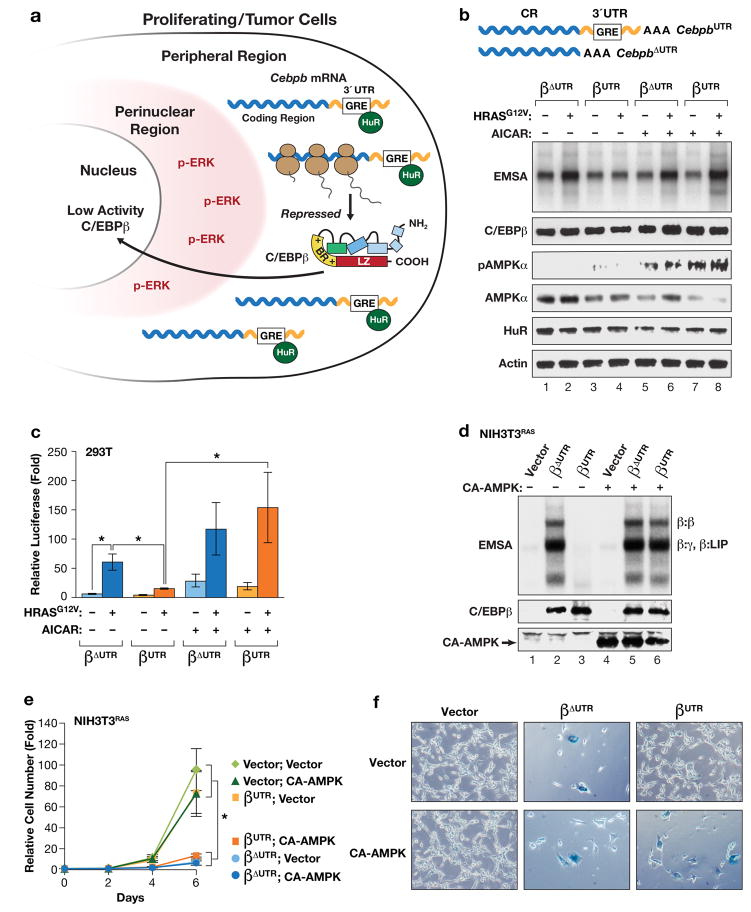
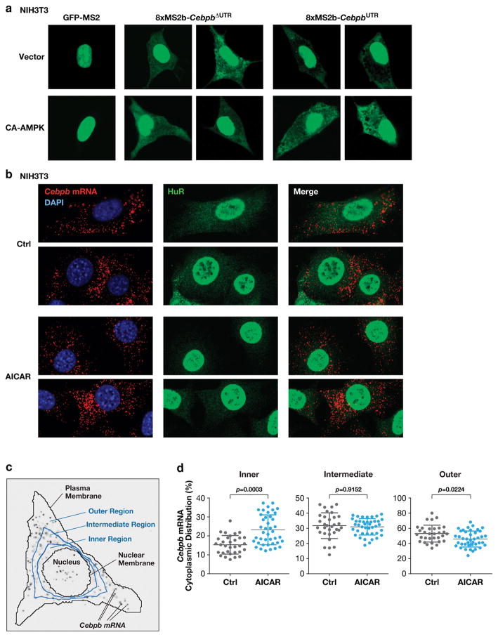
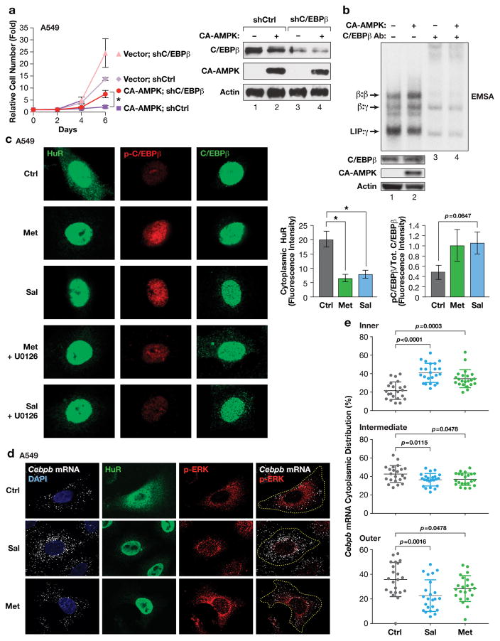
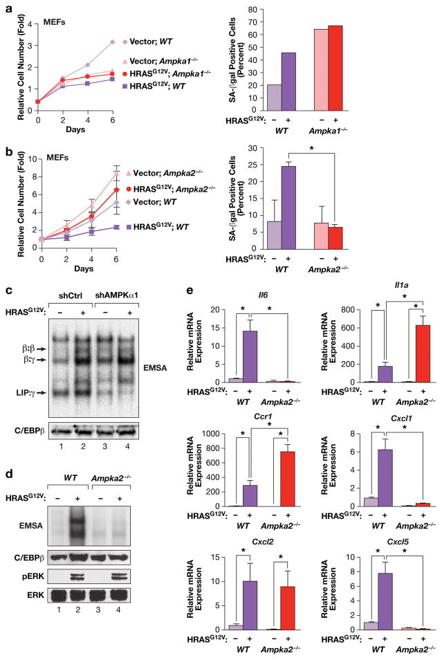
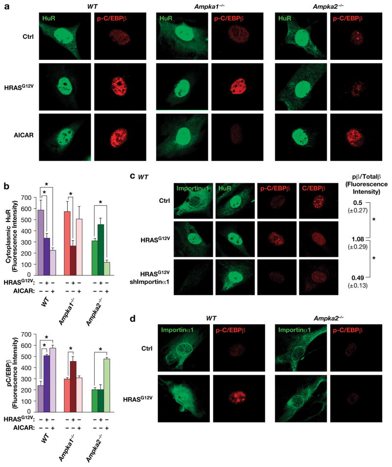
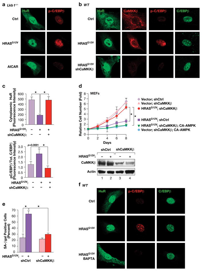
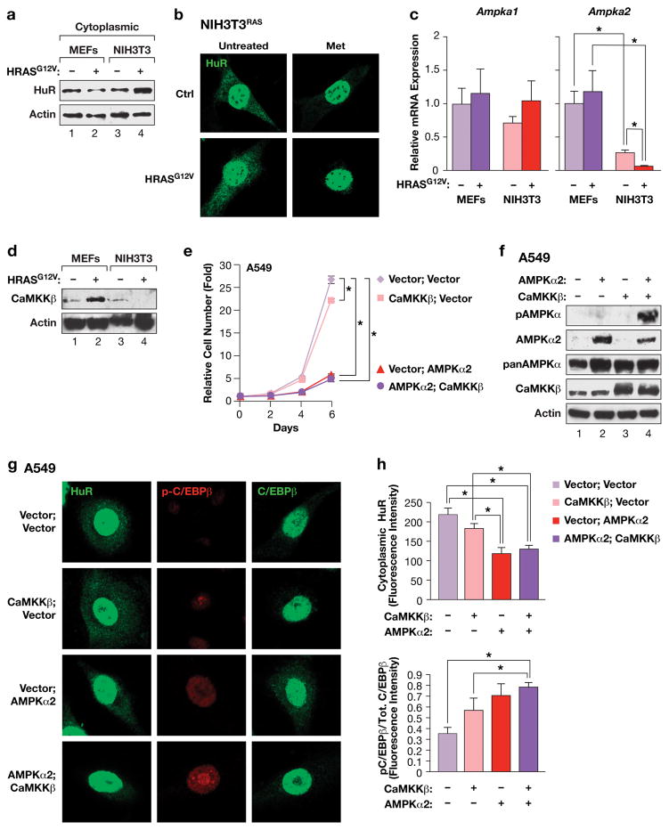
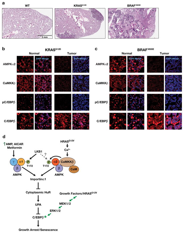
Oncogene-induced senescence (OIS) is an intrinsic tumor suppression mechanism that requires the p53 and RB pathways and post-translational activation of C/EBPβ through the RAS-ERK cascade. We previously reported that in transformed/proliferating cells, C/EBPβ activation is inhibited by G/U-rich elements (GREs) in its 3'UTR. This mechanism, termed "3'UTR regulation of protein activity" (UPA), maintains C/EBPβ in a low-activity state in tumor cells and thus facilitates senescence bypass. Here we show that C/EBPβ UPA is overridden by AMPK signaling. AMPK activators decrease cytoplasmic levels of the GRE binding protein HuR, which is a key UPA component. Reduced cytoplasmic HuR disrupts 3'UTR-mediated trafficking of Cebpb transcripts to the peripheral cytoplasm-a fundamental feature of UPA-thereby stimulating C/EBPβ activation and growth arrest. In primary cells, oncogenic RAS triggers a Ca++-CaMKKβ-AMPKα2-HuR pathway, independent of AMPKα1, that is essential for C/EBPβ activation and OIS. This axis is disrupted in cancer cells through down-regulation of AMPKα2 and CaMKKβ. Thus, CaMKKβ-AMPKα2 signaling constitutes a key tumor suppressor pathway that activates a novel UPA-cancelling mechanism to unmask the cytostatic and pro-senescence functions of C/EBPβ.


Conflict of interest statement

CONFLICT OF INTEREST

The authors declare no conflict of interest.

Figures

Figure 1
Figure 1
AMPK signaling abrogates 3′UTR inhibition of RAS-induced C/EBPβ activation. (a) Model depicting “3′UTR regulation of protein activity” (UPA) in proliferating and transformed cells. The UPA mechanism involves mutually exclusive localization of Cebpb mRNAs (in the peripheral cytoplasm) and the C/EBPβ kinase, activated ERK1/2 (p-ERK) (in the perinuclear cytoplasm). (b, c) The AMPK agonist AICAR overrides UPA to activate C/EBPβ. The effect of AICAR on C/EBPβ DNA binding (b) and transactivation (c) was analyzed in HEK293 cells. Cells were transfected with C/EBPβ constructs containing or lacking the Cebpb 3′UTR (βUTR and βΔUTR, respectively), without or with HRASG12V, and treated with vehicle or 1 mM AICAR for 16 hr prior to harvest. In (b), nuclear extracts normalized for C/EBPβ levels were analyzed by EMSA using a consensus C/EBP probe. The image was cropped to remove the top and bottom (free probe) portions of the gel. In (c), transactivation assays were performed using a C/EBP reporter, 2XC/EBP-Luc. Luciferase activity, normalized to total protein in each lysate, is plotted as fold increase over the reporter alone. n=3; error bars represent S.E.M. Statistical differences between groups were determined by Student’s two-tailed t test; *p<0.05. (d) Expression of a constitutively active AMPKα1 catalytic subunit (CA-AMPK) reverses UPA inhibition of C/EBPβ DNA binding in RAS-transformed NIH3T3 cells. NIH3T3RAS cells, which express low levels of endogenous C/EBPβ, were infected with retroviruses expressing βUTR or βΔUTR, without or with CA-AMPK, and assayed for C/EBPβ DNA binding by EMSA. The various C/EBPβ dimeric complexes are indicated. γ: C/EBPγ; LIP is a truncated translational isoform of C/EBPβ. (e) The same cells were analyzed for proliferation over a 6-day time course. n=3; error bars represent S.E.M. Statistical differences between groups were determined by Student’s t test; *p<0.05. (f) The cells were also stained for the senescence marker, SA-β-Gal. The proportion of SA-β-Gal+ cells in each population is shown in Supplementary Fig. 1c.
Figure 2
Figure 2
AMPK signaling decreases cytoplasmic HuR levels and disrupts peripheral localization of Cebpb transcripts. (a) NIH3T3 cells were transfected with the MS2-GFP-nls reporter alone or together with MS2 binding site-tagged CebpbΔUTR or CebpbUTR vectors, ±CA-AMPK. After 42 hr, cells were analyzed by confocal fluorescence microscopy to visualize cytoplasmic distribution of the transcripts. (b) Localization of endogenous Cebpb mRNA in NIH3T3 cells using RNA FISH. Control and AICAR treated cells were analyzed by FISH using a Cebpb hybridization probe. The cells were also immunostained for HuR. (c) Scheme for quantification of Cebpb mRNA distribution. The cytoplasm was segmented into inner, intermediate, and outer regions by dividing the nuclear-plasma membrane distance radially into thirds. The proportion of the total RNA FISH signal in the three regions was determined for each cell analyzed. (d) Quantitative data from the experiment of panel (b) are shown in the scatter plots. n=32 control cells, 41 AICAR-treated cells. Statistical significance was calculated using Student’s two-tailed t test; *p<0.05.
Figure 3
Figure 3
AMPK signaling activates C/EBPβ in human tumor cells by negatively regulating the HuR-UPA pathway. (a) CA-AMPK induces proliferation arrest in A549 lung adenocarcinoma cells that is partially dependent on C/EBPβ. CA-AMPK was expressed with control or C/EBPβ knockdown vectors and cell proliferation was analyzed over a time course. Levels of CA-AMPK and C/EBPβ were determined by immunoblotting (right panel). n=2 experiments, assayed in triplicate; error bars represent S.E.M. Statistical differences between groups were determined by Student’s t test; *p<0.05. (b) Activated AMPK stimulates C/EBPβ DNA binding and homodimerization in A549 cells. Nuclear extracts from control and CA-AMPK-expressing cells were analyzed by EMSA. C/EBPβ protein levels were normalized in the EMSA reactions so that changes in intrinsic binding activity could be assessed. C/EBPβ-containing complexes were identified by antibody supershift assays (lanes 3–4). (c) The AMPK activators metformin and salicylate induce HuR nuclear translocation and increased phosphorylation on C/EBPβ Thr235 in A549 cells. Right panels show quantification of HuR cytoplasmic intensity and p-C/EBPβ:total C/EBPβ levels in control and drug-treated cells. n=5 cells; statistical significance was calculated using Student’s two-tailed t test; *p<0.05. (d) CEBPB transcripts are excluded from the perinuclear region in A549 cells but become more uniformly distributed following metformin or salicylate treatment. mRNAs were visualized by RNA FISH; the cells were also immunostained for p-ERK and HuR. (e) Cytoplasmic CEBPB mRNA distribution in cells described in panel (d) was quantified as detailed in Fig. 2. n=22 control cells, 23 salicylate-treated cells, 22 metformin-treated cells. Statistical significance was calculated using Student’s two-tailed t test; *p<0.05.
Figure 4
Figure 4
HRASG12V-induced senescence and C/EBPβ activation in MEFs is dependent on AMPKα2. (a) RAS-induced growth arrest and senescence is independent of AMPKα1 but requires AMPKα2. WT and Ampka1−/− MEFs were infected with control or HRASG12V-expressing retroviruses, and cell proliferation (left panel) and senescence (SA-β-Gal staining, right panel) were analyzed. n=1 experiment. b) RAS-induced growth arrest and senescence requires AMPKα2. WT and Ampka2−/− MEFs were infected with control or HRASG12V-expressing retroviruses, and cell proliferation (left panel) and senescence (SA-β-Gal staining, right panel) were analyzed. n=3 experiments for growth curves (assayed in triplicate), n=2 for SA-β-Gal assays; error bars represent S.E.M. Statistical differences were determined by Student’s t test; *p<0.05. (c) C/EBPβ DNA binding is not affected by loss of AMPKα1. AMPKα1 was depleted in WT MEFs by shRNA knockdown, without or with expression of HRASG12V. Nuclear extracts were analyzed by EMSA using a canonical C/EBP site probe. (d) RAS-induced activation of C/EBPβ DNA binding is disrupted in Ampka2−/− MEFs. Nuclear extracts were analyzed by EMSA using a canonical C/EBP site probe. Levels of C/EBPβ, p-ERK and total ERK are also shown. (e) Analysis of SASP gene expression in WT and Ampka2−/− MEFs by qRT-PCR. n=2 biological replicates, each sample assayed in triplicate. Values are averages and error bars represent S.D. Statistical significance was calculated using Student’s two-tailed t test; *p<0.05.
Figure 5
Figure 5
AMPKα2 is required for HuR translocation and C/EBPβ phosphorylation induced by HRASG12V but not AICAR. (a) Control, HRASG12V-expressing and AICAR-treated MEFs (WT, Ampka1−/− and Ampka2−/−) were analyzed by IF staining to assess HuR localization and p-C/EBPβ (Thr188) levels. (b) Cytoplasmic HuR levels and p-C/EBPβ:total C/EBPβ ratios were determined by quantitating images from the experiment of panel (a). n=4 cells; error bars represent S.E.M. Statistical differences were determined by Student’s t test; *p<0.05. (c) The nuclear transport chaperone, Importin α1, undergoes HRASG12V-induced nuclear translocation and is required for HuR shuttling and C/EBPβ activation. WT MEFs were infected with control or HRASG12V-expressing retroviruses, without or with Importin α1 knockdown (shImportin α1). Cells were immunostained for Importin α1, HuR, p-C/EBPβ, and total C/EBPβ. p-C/EBPβ:total C/EBPβ ratios are shown at the right. n=4 cells; error bars represent S.E.M. Statistical differences were determined by Student’s t test; *p<0.05. (d) AMPKα2 is required for nuclear translocation of Importin α1. WT and Ampka2−/− MEFs were analyzed for HRASG12V-induced Importin α1 nuclear re-localization and C/EBPβ phosphorylation (Thr188).
Figure 6
Figure 6
The AMPK kinase CaMKKβ, but not LKB1, controls HRASG12V-induced nuclear translocation of HuR and C/EBPβ activation through a Ca++-dependent pathway. (a) IF staining of HuR and p-C/EBPβ in control and HRASG12V-expressing or AICAR-treated Lkb1−/− MEFs. (b) CaMKKβ depletion prevents HRASG12V-induced activation of the HuR-C/EBPβ pathway. WT MEFs expressing non-specific or shCaMKKβ hairpin RNAs, ± HRASG12V, were analyzed by IF staining for HuR, CaMKKβ, p-C/EBPβ (Thr188) and total C/EBPβ. (c) Quantification of cytoplasmic HuR and p-C/EBPβ:total C/EBPβ ratios from the experiment of panel (b). n=7 cells; error bars represent S.E.M. Statistical differences were determined by Student’s t test; *p<0.05. (d) Proliferation assays were performed using the cells described in panel (b) without or with expression of CA-AMPKα2. n=3; error bars represent S.E.M. Statistical differences at day 8 were determined by Student’s t test; *p<0.05. Lower panel: immunoblot confirming CaMKKβ depletion. (e) Effect of CaMKKβ depletion on RAS-induced senescence (SA-β-Gal positive cells). n=130 cells scored from two independent experiments; error bars represent S.E.M. Statistical differences were determined by Student’s t test; *p<0.05. (f) The Ca++ chelator, BAPTA, blocks HRASG12V-induced HuR nuclear translocation and C/EBPβ phosphorylation. Control and HRASG12V-expressing MEFs were treated with vehicle or 10 μM BAPTA for 2 hr prior to fixation. The cells were immunostained for the indicated proteins.
Figure 7
Figure 7
Proliferation of RAS-transformed cells is associated with elevated cytoplasmic HuR and requires down-regulation of AMPKα2 and CaMKKβ. (a) Immunoblot of cytoplasmic HuR levels in MEFs and NIH3T3 cells, without or with expression of HRASG12V. (b) Immunostaining of HuR in control or HRASG12V-transformed NIH3T3 cells, ± metformin treatment. (c) Ampka1 and Ampka2 mRNA levels in MEFs and NIH3T3 cells, ± HRASG12V. Transcript levels were determined by qRT-PCR. n=3 independent biological replicates, each sample assayed in triplicate; error bars represent S.D. Statistics were calculated using Student’s two tailed t test.; *p<0.05. (d) Immunoblot comparing CaMKKβ levels in MEFs and NIH3T3 cells, without or with expression of HRASG12V. (e) Proliferation of A549 cells over-expressing AMPKα2 and/or CaMKKβ. n=2 independent biological replicates, each time point assayed in triplicate; error bars represent S.E.M. Statistics were determined for day 6 using Student’s two tailed t test.; *p<0.05. (f) Immunoblot showing over-expression of AMPKα2 and CaMKKβ and CaMKKβ-dependent phosphorylation of AMPKα2 in A549 cells. Lysates from the cell populations described in panel (e) were analyzed on blots probed with the indicated antibodies. The anti-p-Thr172 (AMPKα) antibody recognizes modified forms of both AMPKα isoforms. (g) IF imaging of A549 cells expressing AMPKα2 and/or CaMKKβ. Cells were immunostained for HuR, or p-C/EBPβ and total C/EBPβ. (h) Quantitation of cytoplasmic HuR levels and p-C/EBPβ:total C/EBPβ ratios in the cells described in panel g. n=9 cells; error bars represent S.E.M. Statistical differences were determined by Student’s t test; *p<0.05.
Figure 8
Figure 8
AMPKα2, CaMKKβ and p-C/EBPβ levels are markedly decreased in KRASG12D- and BRAFV600E-driven mouse lung tumors. (a) H&E stained lung areas from a normal WT mouse, a KrasLA2/+ mouse containing adenocarcinomas (ADC) and a LSL-BRAFV600E/+ animal bearing multiple adenomas. (b) Normal and lung tumor areas from a 165 day-old KrasLA2/+ mouse; sections were immunostained for AMPKα2, CaMKKβ, p-C/EBPβ (Thr188) and total C/EBPβ. (c) Normal and lung tumor areas from a 190 day-old LSL-BRAFV600E/+ animal (103 days after intratracheal instillation of Ad.Cre virus); sections were immunostained for AMPKα2, CaMKKβ, p-C/EBPβ (Thr188) and total C/EBPβ. (d) Model depicting AMPK-dependent pathways that mediate C/EBPβ activation and senescence in response to energy stress/AMPK agonists or oncogenic RAS. AMPK signaling suppresses the UPA mechanism that inhibits C/EBPβ activation and thus licenses C/EBPβ activation. To become activated, C/EBPβ also requires signaling through the RAS-ERK cascade to induce phosphorylation on Thr188 as well as other modifications,.

Similar articles

Cited by

References

    1. Campisi J. Senescent cells, tumor suppression, and organismal aging: good citizens, bad neighbors. Cell. 2005;120:513–522. - PubMed
    1. Kuilman T, Michaloglou C, Mooi WJ, Peeper DS. The essence of senescence. Genes Dev. 2010;24:2463–2479. - PMC - PubMed
    1. Coppe J-P, Desprez P-Y, Krtolica A, Campisi J. The Senescence-Associated Secretory Phenotype: The Dark Side of Tumor Suppression. Annual Review of Pathology: Mechanisms of Disease. 2010;5:99–118. - PMC - PubMed
    1. Nakajima T, Kinoshita S, Sasagawa T, Sasaki K, Naruto M, Kishimoto T, et al. Phosphorylation at threonine-235 by a ras-dependent mitogen-activated protein kinase cascade is essential for transcription factor NF-IL6. Proc Natl Acad Sci USA. 1993;90:2207–2211. - PMC - PubMed
    1. Lee S, Shuman JD, Guszczynski T, Sakchaisri K, Sebastian T, Copeland TD, et al. RSK-mediated phosphorylation in the C/EBP{beta} leucine zipper regulates DNA binding, dimerization, and growth arrest activity. Mol Cell Biol. 2010;30:2621–2635. - PMC - PubMed

Publication types