. 2018 Jun;37(26):3562-3574.
doi: 10.1038/s41388-018-0191-6. Epub 2018 Mar 22.

HIPK2 and extrachromosomal histone H2B are separately recruited by Aurora-B for cytokinesis

Affiliations

HIPK2 and extrachromosomal histone H2B are separately recruited by Aurora-B for cytokinesis

Laura Monteonofrio et al. Oncogene. 2018 Jun.

Abstract

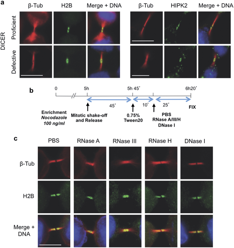
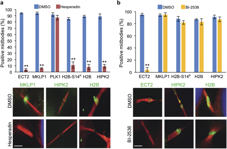
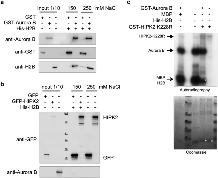
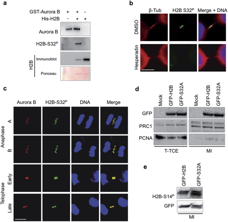
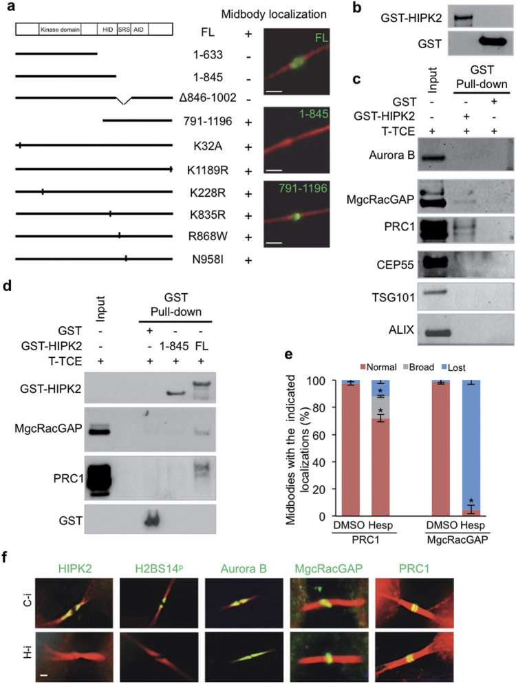
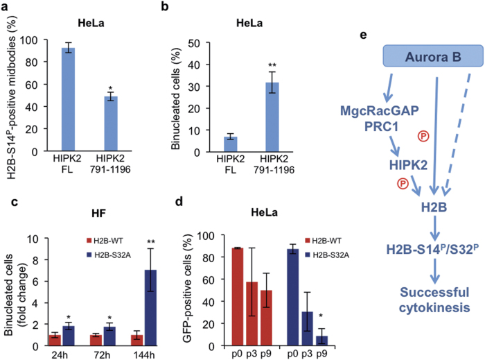
Cytokinesis, the final phase of cell division, is necessary to form two distinct daughter cells with correct distribution of genomic and cytoplasmic materials. Its failure provokes genetically unstable states, such as tetraploidization and polyploidization, which can contribute to tumorigenesis. Aurora-B kinase controls multiple cytokinetic events, from chromosome condensation to abscission when the midbody is severed. We have previously shown that HIPK2, a kinase involved in DNA damage response and development, localizes at the midbody and contributes to abscission by phosphorylating extrachromosomal histone H2B at Ser14. Of relevance, HIPK2-defective cells do not phosphorylate H2B and do not successfully complete cytokinesis leading to accumulation of binucleated cells, chromosomal instability, and increased tumorigenicity. However, how HIPK2 and H2B are recruited to the midbody during cytokinesis is still unknown. Here, we show that regardless of their direct (H2B) and indirect (HIPK2) binding of chromosomal DNA, both H2B and HIPK2 localize at the midbody independently of nucleic acids. Instead, by using mitotic kinase-specific inhibitors in a spatio-temporal regulated manner, we found that Aurora-B kinase activity is required to recruit both HIPK2 and H2B to the midbody. Molecular characterization showed that Aurora-B directly binds and phosphorylates H2B at Ser32 while indirectly recruits HIPK2 through the central spindle components MgcRacGAP and PRC1. Thus, among different cytokinetic functions, Aurora-B separately recruits HIPK2 and H2B to the midbody and these activities contribute to faithful cytokinesis.


Conflict of interest statement

The authors declare that they have no conflict of interest.

Figures

Fig. 1
Fig. 1
H2B and HIPK2 localize at midbody independently of RNA. a Representative images of proliferating HCT116 DICER-proficient and -defective cells analyzed for midbody localization of histone H2B (green; left panels) and HIPK2 (green, right panels). Midbodies were marked with anti-β-tubulin Ab (β-Tub, red); DNA was visualized with Hoechst (blue). b Schematic representation of HeLa cells telophase enrichment with nocodazole, permeabilization, and treatment with RNase A, RNase III, and RNase H to cleave, respectively, single-strand RNA, double-strand RNA, and RNA/DNA hybrid. PBS and DNase I were used as negative controls. c After treatment, cells were fixed and stained with anti-β-tubulin Ab (red) and anti-H2B (green). DNA was marked with Hoechst (blue). Representative images of HeLa cells treated with the indicated enzymes. Each midbody visualized (n > 50 in two independent experiments) was positive for H2B staining. Scale bar is 10 μm
Fig. 2
Fig. 2
The kinase activity of Aurora-B, but not of PLK1, is required for midbody localization of HIPK2 and H2B. a, b Unsynchronized HeLa cells were treated with 100 nM Hesperadin for 80 min or with 1 µM Bi 2536 for 20 min; solvent DMSO was used as control. After treatment, cells were fixed and stained with the indicated Abs (green) in combination with anti-β-tubulin Ab (red). DNA was visualized with Hoechst (blue). The percentages of midbodies positive for the indicated proteins were reported as mean ± SD of three independent experiments. Representative images of indicated staining at the midbody are reported below each chart. **p < 0.001. Scale bar is 1 μm
Fig. 3
Fig. 3
Aurora-B directly binds and phosphorilates H2B. a Recombinant His-H2B was incubated in the presence of recombinant GST-Aurora-B or GST alone at two different salt concentrations. Proteins were detected by Western blotting (WB) with the indicated Abs after GST-pull-down of two independent experiment. b GFP or GFP-HIPK2 were incubated with recombinant GST-Aurora-B at the same conditions described above. Proteins were detected by WB with the indicated Abs after GFP-immunoprecipitation of two independent experiment. c In vitro kinase assays were performed with GST-Aurora-B, as enzymatic source, and His-H2B or GST-HIPK2-K228R as substrates. Myelin basic protein (MBP) was used as positive control. Proteins were separated by SDS-PAGE and radioactivity detected by autoradiography. Coomassie stain was performed as loading control. n = three independent experiment. White asterisks mark nonspecific bands
Fig. 4
Fig. 4
Aurora-B phosphorylates H2B at Ser32. a Cold in vitro kinase assays were performed and H2B-S32P detected using anti-p-Histone H2B-Ser32 Ab. Ponceau staining and immunoblot with anti-H2B Ab were used as loading controls. n = three independent experiment. b Unsynchronized HeLa cells were treated with Hesperadin or DMSO for 80 min and stained with anti-p-Histone H2B-S32 and anti-β-tubulin Abs. Hoechst was used to stain DNA (blue). Representative images of cells at telophase stage are reported. Scale bar is 5 μm. c Representative confocal images of HeLa cells at indicated stages of mitosis and cytokinesis showing colocalization between H2B-S32P (green) and Aurora-B (red) n = four independent experiment. DNA was stained with Red-Dot2 far-red (pseudo-colored blue); scale bar is 10 μm. d, e HeLa cells stably expressing GFP-H2B WT or its derivative GFP-H2B-S32A at passage two after the establishment of stable populations (i.e., 15 days after blastacidin treatment) were enriched in telophase and protein lysates were obtained from TCEs (T-TCE) or after midbody isolation (MI). Untrasfected HeLa cells were used as negative control. The indicated proteins were analyzed by WB. Immunoblots with anti-PRC1 and anti-p-Histone H2B-S14 Abs were used as positive control to verify the quality of midbody isolation. Immunoblot with anti-PCNA Ab was used as control to evaluate nuclear contamination. Representative WBs are shown. In d, samples were loaded on the same gel and processed on the same filter. Blot was vertically cropped to eliminate non-related samples
Fig. 5
Fig. 5
SRS region is essential for HIPK2 midbody localization and interaction with PRC1 and MgcRacGAP central spindle factors. a HeLa cells were transfected with vectors expressing tagged HIPK2-FL or relative derivative mutants. Numbers are referred to the current NCBI RefSeq of HIPK2 (NP_073577.3 and NP_034563.2). Cells were analyzed 24 h post-transfection by IF performed with anti-β-tubulin Ab to mark midbodies (red) in combination with opportune anti-tag Abs to visualize midbody localization of tagged HIPK2 forms. GFP-tagged proteins were visualized by autofluorescence. Midbodies of transfected cells were analyzed for the presence of tagged HIPK2 or relative mutants at the midbody in two independent experiments. Here reported is a schematic representation of the analyzed HIPK2 constructs with their localization at the midbody. Plus (+) indicates midbody localization in >90% of the cells; minus (−) indicates midbody localization in <5% of the cells. Left panel: schematic representation of HIPK2-FL with its domains (HID homeobox Interacting domain, SRS region containing speckle retention sequence, AID auto-inhibitory domain) and its deletion or point mutant forms. Right panels: representative images of midbodies with or without the indicated GFP-HIPK2 forms. Scale bar is 2 μm. b, c GST-HIPK2 and GST alone were produced in H1299 cells, purified by GST pull-down, analyzed by WB b and incubated with an equal amount of T-TCE (0.6 mg) obtained as in Fig. 4d. Proteins bound to GST-HIPK2 and GST were pulled-down and analyzed by WB with the indicated Abs. Representative WBs are shown c; input corresponds to 0.3% of T-TCE. d T-TCE from HeLa cells were obtained and incubated with the indicated GST-fusion proteins as in b,c. The presence of the indicated cytokinetic proteins was assessed by WB. Representative WBs are shown. Input corresponds to 0.3% of the T-TCE. e HeLa cells were treated with Hesperadin as in the Fig. 2. At least 100 midbodies for each condition have been scored and the percentages of midbodies with the indicated localization (i.e., normal, broad, and lost) are reported as mean ± SD of two independent experiments. *p < 0.05. f HeLa cells were transfected with HIPK2-specific stealth siRNAs (H-i) or negative control siRNA (C-i), fixed 4 days after transfection and analyzed by IF with anti-β-tubulin in combination with Abs against the indicated proteins. Scale bar is 1 μm
Fig. 6
Fig. 6
Impairment of Aurora-B-mediated activities on H2B and HIPK2 induces cytokinesis failure. a, b HeLa cells were transfected with vectors carrying the indicated GFP-HIPK2 forms and stained 24 h post-transfection with anti-p-Histone H2B-S14 and Hoechst. The percentages of midbodies positive for H2B-S14P staining in the transfected populations a and the percentages of binucleated cells also in the transfected populations b are reported as mean ± SD of two different experiments. c HF were transfected with vectors carrying GFP-tagged H2B-WT or the non-phosphorylatable H2B-S32A derivative. Cells were stained with anti-β-tubulin Ab and Hoechst. The percentages of binucleated cells in the GFP-positive populations were evaluated at the indicated time-points post-transfection and reported as fold change relative to that of GFP-H2B-WT in two different experiments. *p < 0.05 and **p < 0.001 unpaired t-test. d HeLa cells were transfected with the same vectors as in b and treated with blastacidin 48 h after transfection to select stable transfected cells. The percentage of GFP-positive cells at 2 days (p0) after transfection, or 21 and 39 days after transfection (i.e., at the 3rd and 9th culture passage after blastacidin selection) is reported as mean ± SD of two different experiments. *p < 0.05 unpaired t-test, comparison between percentages of H2B-WT- and H2B-S32A-positive cells at each passage. e Schematic representation of Aurora-B activities on HIPK2 and H2B in cytokinesis. Aurora-B indirectly recruits HIPK2 via MgcRacGAP and PRC1 and directly phosphorylates H2B at Ser32. Additional Aurora-B-dependent event/s, besides phosphorylation on S32, are required for H2B recruitment. Dashed lines indicate still unknown mechanism/s. Phosphorylation of histone H2B at Ser32 by Aurora-B and Ser14 by HIPK2 contribute the successful cytokinesis

Similar articles

Cited by

References

    1. Normand G, King RW. Understanding cytokinesis failure. Ad Exp Med Biol. 2010;676:27–55. doi: 10.1007/978-1-4419-6199-0_3. - DOI - PMC - PubMed
    1. Storchova Z, Kuffer C. The consequences of tetraploidy and aneuploidy. J Cell Sci. 2008;121:3859–66. doi: 10.1242/jcs.039537. - DOI - PubMed
    1. Vitale I, Manic G, Senovilla L, Kroemer G, Galluzzi L. Karyotypic aberrations in oncogenesis and cancer therapy. Trends Cancer. 2015;1:124–35. doi: 10.1016/j.trecan.2015.08.001. - DOI - PubMed
    1. Fededa JP, Gerlich DW. Molecular control of animal cell cytokinesis. Nat Cell Biol. 2012;14:440–7. doi: 10.1038/ncb2482. - DOI - PubMed
    1. Mierzwa B, Gerlich DW. Cytokinetic abscission: molecular mechanisms and temporal control. Dev Cell. 2014;31:525–38. doi: 10.1016/j.devcel.2014.11.006. - DOI - PubMed

Publication types

LinkOut - more resources