

Case Reports
doi: 10.1007/s13193-017-0683-9.
Epub 2017 Aug 23.
Recurrent Bleeding Neck Mass: a Case Report
Affiliations
- PMID: 29563736
- PMCID: PMC5856690
- DOI: 10.1007/s13193-017-0683-9
Item in Clipboard
Case Reports
Recurrent Bleeding Neck Mass: a Case Report
Indian J Surg Oncol.
2018 Mar.
Abstract
An adult male presented to us with a recurrent, large bleeding tumour in the neck. We describe our approach to the patient, whose tumour was labelled as an atypical glomus on final histopathology. They are relatively uncommon in the head neck, and this case report with literature review is expected to add to our knowledge.
Keywords: Atypical glomus; Extradigital glomus.
Figures
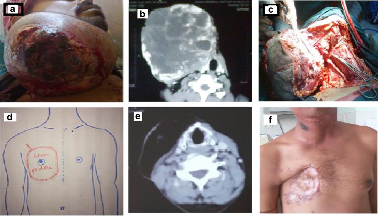
a The neck swelling was 20 × 10 cm, poorly compressible, non-pulsatile boggy mass, with a diffusely bleeding, ulcerated surface, arising from a sessile base from the entire right side of neck. b CECT of the neck showed a well-delineated heterogenously enhancing vascular lesion with dilated vessels supplied by external carotid. The deeper structures appeared normal. c Intraoperative picture showing clamp on external carotid artery. d PMMC markings—our myocutaneous flap was designed with the skin paddle around the nipple, instead of inferior and medial to it, keeping in mind the dimensions of the defect. e CECT neck done 1 year after surgery and RT does not show any recurrence. f Clinically, at 1-year follow-up, well-healed primary and donor sites
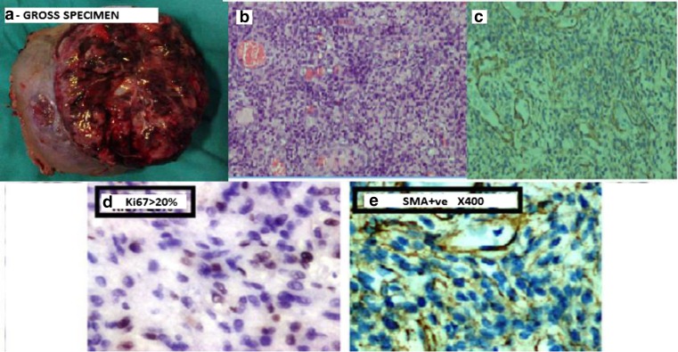
a Gross specimen measured 22 × 19 × 10 cm with areas of necrosis. The cystic spaces varied in size from 0.5 to 1 cm. b Sections showed hypo- and hypercellular areas. The cells are arranged in perivascular pattern and in sheets displaying mild nuclear pleomorphism. Mitosis amounts to 10/10 hpf. (H&E ×100). c The cells were positive for SMA with Ki67 index > 20%. They were negative for CD34, CD31, and desmin. (Slide showing onlySMA positivity). d Sections showing Ki67 positivity> 20%. e ×400 magnification showing SMA positivity
Similar articles
-
A Glomus Tumour of the Elbow: A Case Report and Review of the Literature.Shoulder Elbow. 2014 Jan;6(1):60-2. doi: 10.1111/sae.12041. Epub 2013 Oct 12. Shoulder Elbow. 2014. PMID: 27582912 Free PMC article.
-
Extradigital Glomus Tumor with Atypical Neuritis Presentation.Cureus. 2018 Jun 13;10(6):e2794. doi: 10.7759/cureus.2794. Cureus. 2018. PMID: 30116673 Free PMC article.
-
Extradigital glomus tumour.J Hand Surg Br. 1995 Jun;20(3):409-12. doi: 10.1016/s0266-7681(05)80105-6. J Hand Surg Br. 1995. PMID: 7561424 Review.
-
[Glomus tumor of the thigh: a new case report and literature review].Pan Afr Med J. 2017 Sep 26;28:73. doi: 10.11604/pamj.2017.28.73.12827. eCollection 2017. Pan Afr Med J. 2017. PMID: 29255543 Free PMC article. Review. French.
-
Extradigital Symplastic Glomus Tumor of the Hand: Report of 2 Cases and Literature Review.Am J Dermatopathol. 2015 Jul;37(7):560-2. doi: 10.1097/DAD.0000000000000132. Am J Dermatopathol. 2015. PMID: 25051107 Review.
References
-
- Enzinger FM, Weiss SW. Perivascular tumors. Enzinger and Weiss’s Soft tissue tumors. 4. Maryland Heights: Mosby; 2001. pp. 985–1035.
-
- Lee D-W, Yang J-H, Chang S, Won C-H, Lee M-W, Choi J-H, Moon K-C Clinical and pathological characteristics of extradigital and digital glomus tumours: a retrospective comparative study. J Eur Acad Dermatol Venereol 25:1392–1397. doi:10.1111/j.1468-3083.2011.03979.x - PubMed
Publication types
LinkOut - more resources
Full Text Sources
Other Literature Sources