

DNA nanoparticles are safe and nontoxic in non-human primate eyes
- PMID: 29563793
- PMCID: PMC5849385
- DOI: 10.2147/IJN.S157000
DNA nanoparticles are safe and nontoxic in non-human primate eyes
Abstract
Introduction: DNA nanoparticles (NPs) comprising polylysine conjugated to polyethylene glycol efficiently target murine photoreceptors and the retinal pigment epithelium (RPE) and lead to long-term phenotypic improvement in models of retinal degeneration. Advancing this technology requires testing in a large animal model, particularly with regard to safety. So, herein we evaluate NPs in non-human primates (baboon).
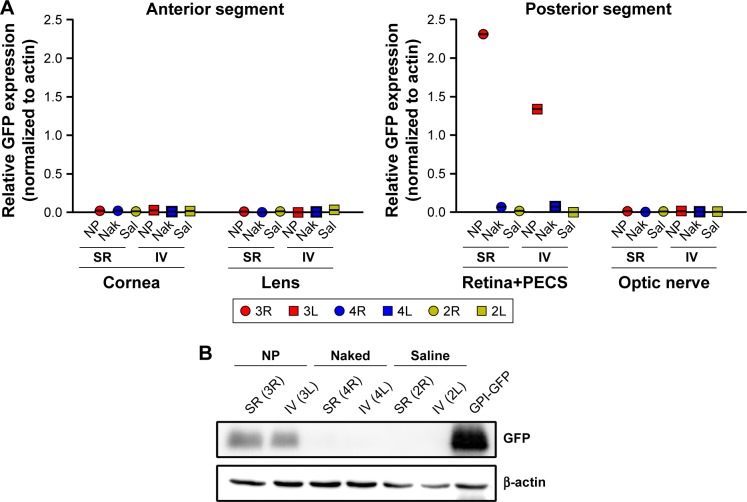
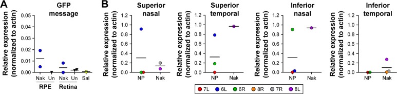
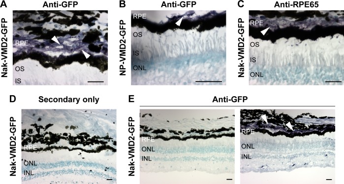
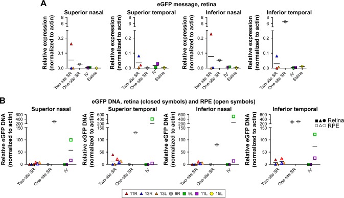
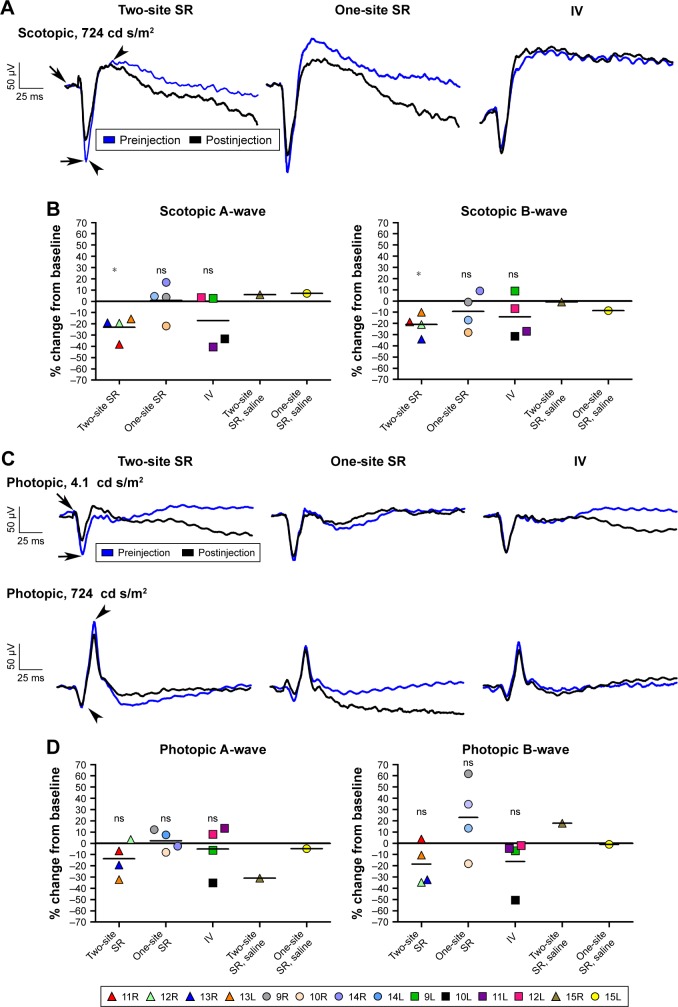
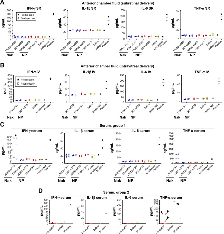
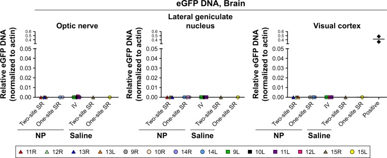
Methods and results: NPs with plasmids carrying GFP and a ubiquitous, RPE-specific, or photoreceptor-specific promoter were delivered by either subretinal or intravitreal injection. We detected GFP message and protein in the retina/RPE from eyes dosed with NPs carrying ubiquitously expressed and RPE-specific vectors, and GFP message in eyes injected with NPs carrying photoreceptor-specific vectors. Importantly, we observed NP DNA in the retina/RPE following intravitreal injection, indicating the inner limiting membrane does not prevent NP diffusion into the outer retina. We did not observe any adverse events in any baboon, and there were no NP-associated changes in retinal function. Furthermore, no systemic or local inflammatory reaction to the vectors/injections was observed, and no NP DNA was found outside the eye.
Conclusion: Taken together with the well-established rodent safety and efficacy data, these findings suggest that DNA NPs may be a safe and potentially clinically viable nonviral ocular therapy platform for retinal diseases.
Keywords: DNA nanoparticles; baboon; gene therapy; non-human primate; nonviral gene transfer; ocular gene transfer; safety.
Conflict of interest statement
Disclosure MJC is an employee of Copernicus Therapeutics and owns stock in the company. The authors report no other conflicts of interest in this work.
Figures
Similar articles
-
Synthesis and characterization of glycol chitosan DNA nanoparticles for retinal gene delivery.ChemMedChem. 2014 Jan;9(1):189-96. doi: 10.1002/cmdc.201300371. Epub 2013 Nov 7. ChemMedChem. 2014. PMID: 24203490 Free PMC article.
-
Nanoparticle-mediated gene transfer specific to retinal pigment epithelial cells.Biomaterials. 2011 Dec;32(35):9483-93. doi: 10.1016/j.biomaterials.2011.08.062. Epub 2011 Sep 1. Biomaterials. 2011. PMID: 21885113 Free PMC article.
-
Self-complementary AAV5 vector facilitates quicker transgene expression in photoreceptor and retinal pigment epithelial cells of normal mouse.Exp Eye Res. 2010 May;90(5):546-54. doi: 10.1016/j.exer.2010.01.011. Epub 2010 Feb 4. Exp Eye Res. 2010. PMID: 20138034 Free PMC article.
-
Suprachoroidal Delivery of Viral and Nonviral Gene Therapy for Retinal Diseases.J Ocul Pharmacol Ther. 2020 Jul/Aug;36(6):384-392. doi: 10.1089/jop.2019.0126. Epub 2020 Apr 7. J Ocul Pharmacol Ther. 2020. PMID: 32255727 Free PMC article. Review.
-
[Retinal pigment epithelial cell transplantation: perspective].Nippon Ganka Gakkai Zasshi. 1996 Dec;100(12):982-1006. Nippon Ganka Gakkai Zasshi. 1996. PMID: 9022310 Review. Japanese.
Cited by
-
Gene Therapy to the Retina and the Cochlea.Front Neurosci. 2021 Mar 17;15:652215. doi: 10.3389/fnins.2021.652215. eCollection 2021. Front Neurosci. 2021. PMID: 33815052 Free PMC article. Review.
-
Lipid nanoparticles for delivery of messenger RNA to the back of the eye.J Control Release. 2019 Jun 10;303:91-100. doi: 10.1016/j.jconrel.2019.04.015. Epub 2019 Apr 12. J Control Release. 2019. PMID: 30986436 Free PMC article.
-
In Silico CRISPR-Cas-Mediated Base Editing Strategies for Early-Onset, Severe Cone-Rod Retinal Degeneration in Three Crumbs homolog 1 Patients, including the Novel Variant c.2833G>A.Genes (Basel). 2024 May 15;15(5):625. doi: 10.3390/genes15050625. Genes (Basel). 2024. PMID: 38790254 Free PMC article.
-
Research Models and Gene Augmentation Therapy for CRB1 Retinal Dystrophies.Front Neurosci. 2020 Aug 14;14:860. doi: 10.3389/fnins.2020.00860. eCollection 2020. Front Neurosci. 2020. PMID: 32922261 Free PMC article. Review.
-
The future of retinal gene therapy: evolving from subretinal to intravitreal vector delivery.Neural Regen Res. 2021 Sep;16(9):1751-1759. doi: 10.4103/1673-5374.306063. Neural Regen Res. 2021. PMID: 33510064 Free PMC article. Review.
References
-
- Lapuerta P, Schein SJ. A four-surface schematic eye of macaque monkey obtained by an optical method. Vision Res. 1995;35:2245–2254. - PubMed
-
- Panda-Jonas S, Jonas JB, Jakobczyk M, et al. Retinal photoreceptor count, retinal surface area, and optic disc size in normal human eyes. Ophthalmology. 1994;101:519–523. - PubMed
-
- Cai X, Cooper M, Naash M. Effective gene transfer for leber congenital amaurosis with compacted Dna nanoparticle. Invest Ophthalmol Vis Sci. 2009;50:1737.
MeSH terms
Substances
Grants and funding
LinkOut - more resources
Full Text Sources
Other Literature Sources
Medical
Miscellaneous
