. 2018 Mar 6:9:83.
doi: 10.3389/fphys.2018.00083. eCollection 2018.

Soluble Fms-Like Tyrosine Kinase-1 Alters Cellular Metabolism and Mitochondrial Bioenergetics in Preeclampsia

Affiliations

Soluble Fms-Like Tyrosine Kinase-1 Alters Cellular Metabolism and Mitochondrial Bioenergetics in Preeclampsia

Lissette C Sánchez-Aranguren et al. Front Physiol. .

Abstract

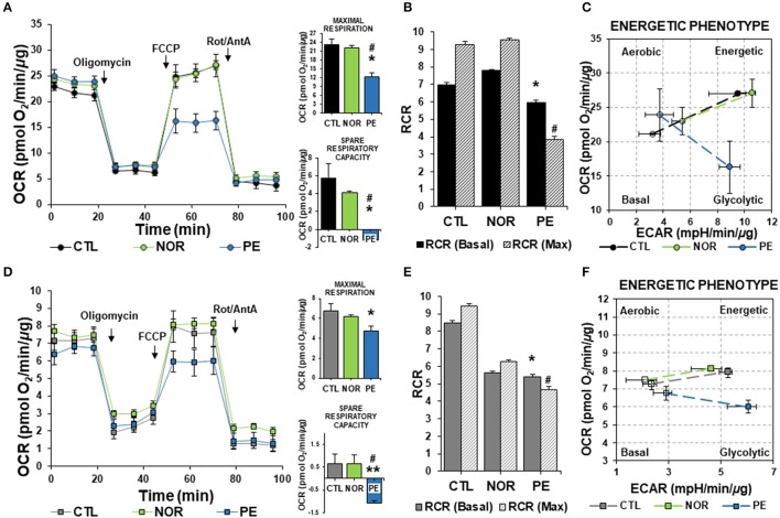
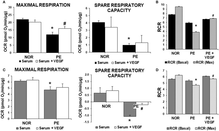
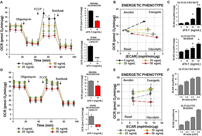
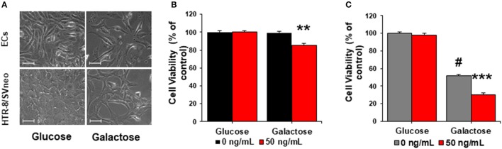
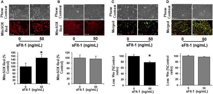
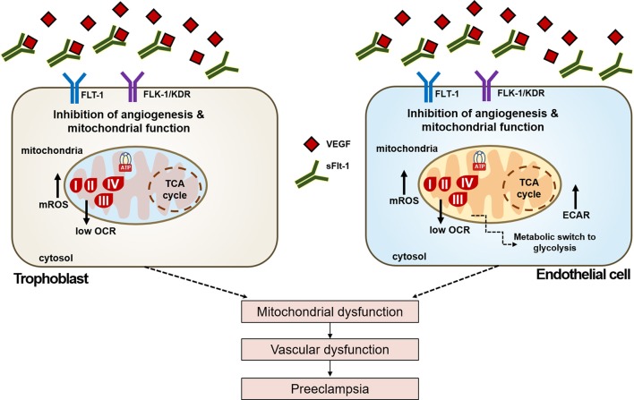
Preeclampsia is a maternal hypertensive disorder that affects up to 1 out of 12 pregnancies worldwide. It is characterized by proteinuria, endothelial dysfunction, and elevated levels of the soluble form of the vascular endothelial growth factor receptor-1 (VEGFR-1, known as sFlt-1). sFlt-1 effects are mediated in part by decreasing VEGF signaling. The direct effects of sFlt-1 on cellular metabolism and bioenergetics in preeclampsia, have not been established. The goal of this study was to evaluate whether sFlt-1 causes mitochondrial dysfunction leading to disruption of normal functioning in endothelial and placental cells in preeclampsia. Endothelial cells (ECs) and first-trimester trophoblast (HTR-8/SVneo) were treated with serum from preeclamptic women rich in sFlt-1 or with the recombinant protein. sFlt-1, dose-dependently inhibited ECs respiration and acidification rates indicating a metabolic phenotype switch enhancing glycolytic flux. HTR-8/SVneo displayed a strong basal glycolytic metabolism, remaining less sensitive to sFlt-1-induced mitochondrial impairment. Moreover, results obtained in ECs exposed to serum from preeclamptic subjects demonstrated that increased sFlt-1 leads to metabolic perturbations accountable for mitochondrial dysfunction observed in preeclampsia. sFlt-1 exacerbated mitochondrial reactive oxygen species (ROS) formation and mitochondrial membrane potential dissipation in ECs and trophoblasts exposed to serum from preeclamptic women. Forcing oxidative metabolism by culturing cells in galactose media, further sensitized cells to sFlt-1. This approach let us establish that sFlt-1 targets mitochondrial function in ECs. Effects of sFlt-1 on HTR-8/SVneo cells metabolism were amplified in galactose, demonstrating that sFlt-1 only target cells that rely mainly on oxidative metabolism. Together, our results establish the early metabolic perturbations induced by sFlt-1 and the resulting endothelial and mitochondrial dysfunction in preeclampsia.

Keywords: endothelial dysfunction; metabolism; mitochondrial dysfunction; oxidative stress and metabolic perturbations; preeclampsia; sFlt-1.


Figures

Figure 1
Figure 1
Bioenergetics profile of cells treated with serum from PE women. ECs and HTR-8/SVneo cells were treated with 2% serum from NOR, CTL, and PE women for 24 h. (A) Basal oxygen consumption rates (OCR), maximal respiration (OCR after FCCP administration) and spare respiratory capacity (Difference between basal and maximal OCR), (B) Ratios of basal and maximal (Max) respiratory control (RCR) (State 3/state 4), (C) Energy phenotype map of basal and maximal OCR and extracellular acidification rates (ECAR), measured in ECs. (D–F), are the same experiments as in (A–C), respectively, but evaluated in HTR-8/SVneo cells. Data are presented as means ± SEM. (n = 5). In (A,D): *P < 0.05, **P < 0.01 vs. CTL, #P < 0.05, vs. NOR. In (B,E): *P < 0.05, vs. basal CTL RCR, #P < 0.05, vs. CTL maximal RCR. ANOVA (Bonferroni's post-hoc test).
Figure 2
Figure 2
Role of VEGF in mitochondrial bioenergetics in preeclampsia. ECs and HTR-8/SVneo cells were treated with 2% serum from NOR, CTL, and PE women with and without exogenous VEGF (20 ng/mL) for 24 h. (A) Maximal respiration (OCR after FCCP administration) and spare respiratory capacity (Difference between basal and maximal OCR) in endothelial cells, (B) Basal and maximal (Max) RCR, (C,D) are the same experiments as in (A,B), respectively, but evaluated in HTR-8/SVneo cells. Data are presented as means ± SEM. (n = 5). In (A,C): *P < 0.05, vs. NOR, #P < 0.05, vs. cells exposed to PE serum alone. In (B,D): *P < 0.05, vs. NOR maximal RCR, #P < 0.05, vs. PE maximal RCR. ANOVA (Bonferroni's post hoc test).
Figure 3
Figure 3
VEGF restores mitochondrial function and abrogates ROS in preeclampsia. ECs and HTR-8/SVneo cells were treated with 2% serum from NOR, CTL, and PE women with and without exogenous VEGF (20 ng/mL) for 24 h. (A) Mitochondrial ROS (mtROS) determinations by fluorescent microscopy using MitoSOX Red fluorescent probe in ECs. (B) As in (A) but with trophoblasts cells. (C) PE serum dissipated the mitochondrial membrane potential (Ψm) measured by fluorescent microscopy using JC-1 fluorescent dye in ECs and (D) trophoblasts. VEGF (20 ng/mL) restored the mitochondrial membrane potential to CTL levels in trophoblasts. Scale: 100 μm. Data are presented as means ± SEM. (n = 3), *P < 0.05, **P < 0.01, ***P < 0.001, vs. CTL exposed cells. #P < 0.05, vs. PE serum exposed cells, ANOVA (Bonferroni's post hoc test).
Figure 4
Figure 4
sFlt-1 induces a metabolic phenotype switch to glycolysis in ECs but not in trophoblasts. ECs and HTR-8/SVneo cells were exposed to 0, 10, 25, and 50 ng/mL of rh-sFlt-1 for 24 h. (A) Oxygen consumption rates (OCR), maximal respiration (OCR after FCCP administration) and spare respiratory capacity (Difference between basal and maximal OCR), (B) Energetic phenotype map, basal and maximal OCR and extracellular acidification rates (ECAR), (C) ECAR determinations of glycolysis rate and glycolytic reserve, evaluated in endothelial cells. (D-F), are the same experiments as in (A–C), respectively, but evaluated in HTR-8/SVneo cells. Data is presented as means ± SEM. (n = 5), *P < 0.05, **P < 0.01, ***P < 0.001 vs. untreated controls, In (A,D): Student T-test. In (C,F): ANOVA (Bonferroni's post hoc test).
Figure 5
Figure 5
sFlt-1 acts as a mitochondrial bioenergetics disruptor in preeclampsia. (A) Morphological changes in endothelial cells (ECs) and trophoblasts cultured in glucose and galactose media (40X). (B) Cell viability of ECs and (C) trophoblasts cultured in glucose and galactose media and exposed to exogenous 50 ng/mL of sFlt-1 during 24 h. Scale: 100 μm. Data are presented as means ± SEM. (n = 3), **P < 0.01, ***P < 0.001, vs. galactose exposed cells. #P < 0.05, vs. glucose-exposed cells. Student T-test.
Figure 6
Figure 6
sFlt-1 induces mitochondrial dysfunction in vitro. (A) Mitochondrial ROS (mtROS) determinations by fluorescent microscopy using MitoSOX Red fluorescent probe demonstrate that sFlt-1 significantly induced mtROS formation in endothelial cells (ECs), while these effects were not observed in (B) trophoblasts. (C) sFlt-1 dissipated the mitochondrial membrane potential (Ψm) measured by fluorescent microscopy using JC-1 fluorescent probe in ECs, but not in (D) trophoblasts. Cells were exposed to sFlt-1 (50 ng/mL) for 24 h. Data is presented as means ± SEM. (n = 3), *P < 0.05 vs. untreated controls. Student T-test.
Figure 7
Figure 7
Schematic view of the effects of sFlt-1 dysregulation over cellular metabolism and bioenergetics in PE. Dysregulated VEGF signaling due to up-regulation of sFlt-1 levels in preeclampsia leads to reduced activation of VEGF receptors Flt-1 and Flk-1/KDR, respectively. Effects of dysregulated VEGF bioavailability affect mitochondrial oxygen consumption (OCR), inducing a metabolic phenotype switch enhancing glycolytic response (ECAR) in endothelial cells, but not in trophoblasts. sFlt-1 due to loss of mitochondrial bioenergetics increase oxidative stress in mitochondria (mtROS). Together, these events lead to mitochondrial dysfunction, that would result in vascular dysfunction and the onset of preeclampsia.

References

    1. American College Obstetricians Gynecologist and Task Force on Hypertension in Pregnancy (2013). Hypertension in pregnancy. Report of the American College of Obstetricians and Gynecologists' Task Force on Hypertension in Pregnancy. Obstet. Gynecol. 122, 1122–1131. 10.1097/01.AOG.0000437382.03963.88 - DOI - PubMed
    1. Benard G., Faustin B., Passerieux E., Galinier A., Rocher C., Bellance N., et al. . (2006). Physiological diversity of mitochondrial oxidative phosphorylation. Am. J. Physiol. Cell Physiol. 291, C1172–C1182. 10.1152/ajpcell.00195.2006 - DOI - PubMed
    1. Brand M. D., Nicholls D. G. (2011). Assessing mitochondrial dysfunction in cells. Biochem. J. 435, 297–312. 10.1042/BJ20110162 - DOI - PMC - PubMed
    1. Chaiworapongsa T., Chaemsaithong P., Yeo L., Romero R. (2014). Pre-eclampsia part 1: current understanding of its pathophysiology. Nat. Rev. Nephrol. 10, 466–480. 10.1038/nrneph.2014.102 - DOI - PMC - PubMed
    1. DeBerardinis R. J., Thompson C. B. (2012). Cellular metabolism and disease: what do metabolic outliers teach us? Cell 148, 1132–1144. 10.1016/j.cell.2012.02.032 - DOI - PMC - PubMed

LinkOut - more resources