. 2018 Mar 7:9:342.
doi: 10.3389/fmicb.2018.00342. eCollection 2018.

A Systematic Evaluation of the Two-Component Systems Network Reveals That ArlRS Is a Key Regulator of Catheter Colonization by Staphylococcus aureus

Affiliations

A Systematic Evaluation of the Two-Component Systems Network Reveals That ArlRS Is a Key Regulator of Catheter Colonization by Staphylococcus aureus

Saioa Burgui et al. Front Microbiol. .

Abstract

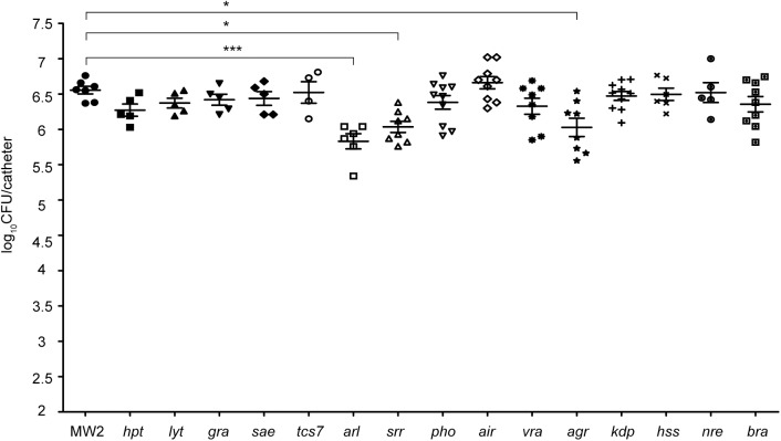
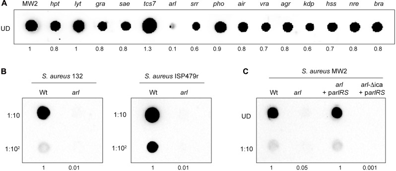
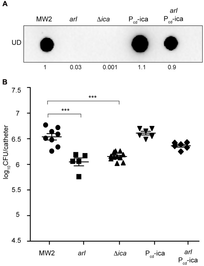
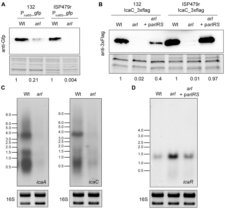
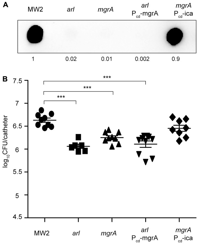
Two-component systems (TCS) are modular signal transduction pathways that allow cells to adapt to prevailing environmental conditions by modifying cellular physiology. Staphylococcus aureus has 16 TCSs to adapt to the diverse microenvironments encountered during its life cycle, including host tissues and implanted medical devices. S. aureus is particularly prone to cause infections associated to medical devices, whose surfaces coated by serum proteins constitute a particular environment. Identification of the TCSs involved in the adaptation of S. aureus to colonize and survive on the surface of implanted devices remains largely unexplored. Here, using an in vivo catheter infection model and a collection of mutants in each non-essential TCS of S. aureus, we investigated the requirement of each TCS for colonizing the implanted catheter. Among the 15 mutants in non-essential TCSs, the arl mutant exhibited the strongest deficiency in the capacity to colonize implanted catheters. Moreover, the arl mutant was the only one presenting a major deficit in PNAG production, the main exopolysaccharide of the S. aureus biofilm matrix whose synthesis is mediated by the icaADBC locus. Regulation of PNAG synthesis by ArlRS occurred through repression of IcaR, a transcriptional repressor of icaADBC operon expression. Deficiency in catheter colonization was restored when the arl mutant was complemented with the icaADBC operon. MgrA, a global transcriptional regulator downstream ArlRS that accounts for a large part of the arlRS regulon, was unable to restore PNAG expression and catheter colonization deficiency of the arlRS mutant. These findings indicate that ArlRS is the key TCS to biofilm formation on the surface of implanted catheters and that activation of PNAG exopolysaccharide production is, among the many traits controlled by the ArlRS system, a major contributor to catheter colonization.

Keywords: PNAG; Staphylococcus aureus; arlRS; biofilm; implants; two-component systems.


Figures

FIGURE 1
FIGURE 1
Schematic illustrating the murine model of catheter-associated biofilm formation. (A) ICR female mice were anesthetized by isoflurane and the skin was cleansed with alcohol prior to surgery. The bottom of 19 mm catheters (Introcan Safety 24G; B. Braun) was cut right before insertion (dotted line). (B) Then, two catheters were inserted into the subcutaneous interscapular space of each mice and 100 μl of the bacterial suspension containing 107 CFU of the strain under study were injected through the Introcan Devices into the catheters. In all the experiments, five mice were used for each strain under study, so that a total of ten catheters were inoculated with each strain. (C) Introcan Devices were carefully pulled out from mice and wounds were closured with the tissue adhesive Histoacryl® (B. Braun) so that catheters remained inside for 5 days. Note that although closuring the wounds, some catheters were naturally pulled out from mice during the course of experiments. (D) Mice were euthanized, catheters were aseptically removed, placed in a sterile microcentrifuge tube containing 1 ml of PBS and vigorously vortexed for 3 min to remove adherent bacteria. (E) Bacteria were enumerated by viable plate counts.
FIGURE 2
FIGURE 2
Systematic analysis of the contribution of the TCS signaling system to catheter colonization. Comparison of catheter colonization capacity of the wild type strain (MW2) and single mutants in each non-essential TCS. Bacteria were not detectable in control catheters that had been inoculated with PBS (detection limit 100 CFU/catheter). Note that although a total of ten catheters were inoculated with each strain, a variable number of catheters were recovered in each group due to natural catheter expulsion from mice during the course of the experiment. The plots display values obtained from individual catheters and the mean is represented by horizontal bars. Statistical significance was determined with one-way ANOVA followed by Tukey’s multiple comparison test comparing to the WT strain. P < 0.05, ∗∗∗P < 0.001.
FIGURE 3
FIGURE 3
Staphylococcus aureus arlRS mutants do not synthesize PNAG. (A) Dot blot analysis of the PNAG exopolysaccharide synthesized by the wild type MW2 strain and the collection of single mutants in each non-essential TCS. (B) Dot blot analysis of PNAG synthesized by the wild type 132 and ISP479r strains and their corresponding arl mutants. (C) Dot blot analysis of PNAG produced by S. aureus MW2, a single arl and double arlica mutant strains complemented with plasmid parlRS, which overexpresses the arlRS genes under the expression of their own promoter. In all cases, samples were analyzed after 16 h of static incubation, at 37°C, in TSB (MW2 and derivatives), TSB NaCl (132) and TSBg (ISP479r). Serial dilutions (1/10) of the samples were spotted onto nitrocellulose membranes and PNAG production was detected with specific anti PIA/PNAG antibodies. UD; undiluted sample. Numbers below the image show relative dot quantification according to densitometry analysis performed with ImageJ (http://rsbweb.nih.gov/ij/).
FIGURE 4
FIGURE 4
Overexpression of the icaADBC operon in an arl mutant restores catheter colonization. (A) Dot blot analysis of the PNAG exopolysaccharide synthesized by the wild type MW2, arl, Δica and the wild type and arl mutant that overproduce PNAG through the chromosomal expression of the icaADBC operon under the Pcad promoter (Pcd_ica and arl Pcd_ica, respectively). Samples were analyzed after 16 h of static incubation, at 37°C, in TSB media. Samples were spotted onto nitrocellulose membranes and PNAG production was detected with specific anti PIA/PNAG antibodies. Numbers below the image show relative dot quantification according to densitometry analysis performed with ImageJ (http://rsbweb.nih.gov/ij/). UD; undiluted sample. (B) Comparison of catheter colonization capacity of the strains shown in (A). Bacteria were not detectable in control catheters that had been inoculated with PBS (detection limit 100 CFU/catheter). Note that although a total of ten catheters were inoculated with each strain, a variable number of catheters were recovered in each group due to natural catheter expulsion from mice during the course of the experiment. The plots display values obtained from individual catheters and the mean is represented by horizontal bars. Statistical significance was determined with one-way ANOVA followed by Tukey’s multiple comparison test comparing to the WT strain. ∗∗∗P < 0.001.
FIGURE 5
FIGURE 5
ArlRS transcriptionally activates icaADBC operon expression in S. aureus. (A) A representative Western blot showing GFP protein levels expressed from S. aureus 132 and ISP479r wild type strains and their corresponding arl mutants harboring plasmid Pica(BS)-gfp. The GFP protein was detected with commercial anti-GFP antibodies. A stain-free gel portion is shown as a loading control. Numbers below the image show relative band quantification according to densitometry analysis performed with ImageJ (http://rsbweb.nih.gov/ij/). (B) Representative Northern blots showing icaA and icaC mRNA of S. aureus 132 wild type and arl mutant strains grown at 37°C until exponential phase (OD600 nm = 0.8). Lower panels show 16S ribosome band stained with RedSafe Nucleic Acid Staining Solution as loading control. (C) A representative Western blot showing IcaC protein levels of the wild type 132 and ISP479r strains, arl mutants and arl mutants complemented with a plasmid expressing the arlRS genes (arl parlRS). The 3XFlag tagged IcaC protein was detected with commercial anti-3XFlag antibodies. A stain-free gel portion is shown as a loading control. Numbers below the image show relative band quantification according to densitometry analysis performed with ImageJ (http://rsbweb.nih.gov/ij/). (D) A representative Northern blot showing icaR mRNA of S. aureus 132 wild type strain, arl mutant and arl mutant complemented with a plasmid expressing the arlRS genes (arl parlRS) grown at 37°C until exponential phase (OD600 nm = 0.8). Lower panels show 16S ribosome band stained with RedSafe Nucleic Acid Staining Solution as loading control.
FIGURE 6
FIGURE 6
MgrA is unable to restore catheter colonization of an arl mutant. (A) Dot blot analysis of the PNAG exopolysaccharide synthesized by the wild type MW2, arl, mgrA, arl that overproduces MgrA through the chromosomal expression of the mgrA gene under the Pcad promoter (arl Pcd_mgrA), and mgrA that overproduces PNAG through the chromosomal expression of the icaADBC operon under the Pcad promoter (mgrA Pcd_ica). Samples were analyzed after 16 h of static incubation, at 37°C, in TSB media. Samples were spotted onto nitrocellulose membranes and PNAG production was detected with specific anti PIA/PNAG antibodies. Numbers below the image show relative dot quantification according to densitometry analysis performed with ImageJ (http://rsbweb.nih.gov/ij/). UD; undiluted sample. (B) Comparison of catheter colonization capacity of S. aureus strains shown in (A). Bacteria were not detectable in control catheters that had been inoculated with PBS (detection limit 100 CFU/catheter). Note that although a total of 10 catheters were inoculated with each strain, a variable number of catheters were recovered in each group due to natural catheter expulsion from mice during the course of the experiment. The plots display values obtained from individual catheters and the mean is represented by horizontal bars. Statistical significance was determined with one-way ANOVA followed by Tukey’s multiple comparison test comparing to the WT strain. ∗∗∗P < 0.001.

Similar articles

Cited by

References

    1. Alanidis C., Dejong P. J. (1990). Ligation-independent cloning of PCR products (LIC-PCR). Nucleic Acids Res. 18 6069–6074. 10.1093/nar/18.20.6069 - DOI - PMC - PubMed
    1. Arnaud M., Chastanet A., Débarbouillé M. (2004). New vector for efficient allelic replacement in naturally nontransformable, low-GC-content, gram-positive bacteria. Appl. Environ. Microbiol. 70 6887–6891. 10.1128/AEM.70.11.6887-6891.2004 - DOI - PMC - PubMed
    1. Briandet R., Herry J. M., Bellon-Fontaine M. N. (2001). Determination of the van der Waals, electron donor and electron acceptor surface tension components of static Gram-positive microbial biofilms. Colloids Surf. B Biointerfaces 21 299–310. 10.1016/S0927-7765(00)00213-7 - DOI - PubMed
    1. Cerca N., Pier G. B., Vilanova M., Oliveira R., Azeredo J. (2005). Quantitative analysis of adhesion and biofilm formation on hydrophilic and hydrophobic surfaces of clinical isolates of Staphylococcus epidermidis. Res. Microbiol. 156 506–514. 10.1016/j.resmic.2005.01.007 - DOI - PMC - PubMed
    1. Charpentier E., Anton A. I., Barry P., Alfonso B., Fang Y., Novick R. P. (2004). Novel cassette-based shuttle vector system for gram-positive bacteria. Appl. Environ. Microbiol. 70 6076–6085. 10.1128/AEM.70.10.6076-6085.2004 - DOI - PMC - PubMed

LinkOut - more resources