. 2018 Mar 7:9:392.
doi: 10.3389/fmicb.2018.00392. eCollection 2018.

Chronic Inflammatory Microenvironment in Epidermodysplasia Verruciformis Skin Lesions: Role of the Synergism Between HPV8 E2 and C/EBPβ to Induce Pro-Inflammatory S100A8/A9 Proteins

Affiliations

Chronic Inflammatory Microenvironment in Epidermodysplasia Verruciformis Skin Lesions: Role of the Synergism Between HPV8 E2 and C/EBPβ to Induce Pro-Inflammatory S100A8/A9 Proteins

Marta Podgórska et al. Front Microbiol. .

Abstract

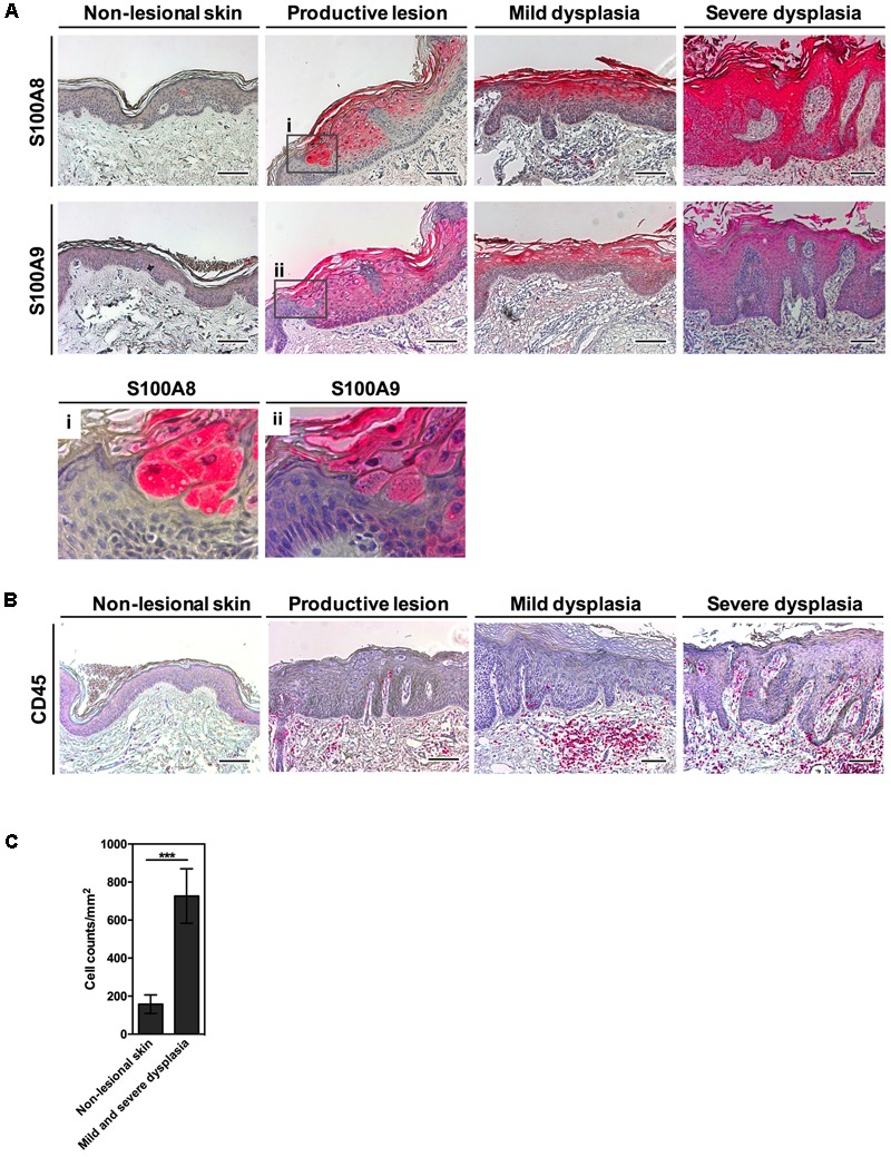
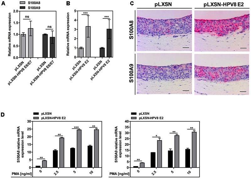
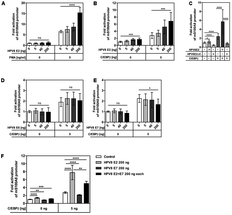
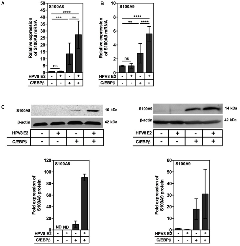
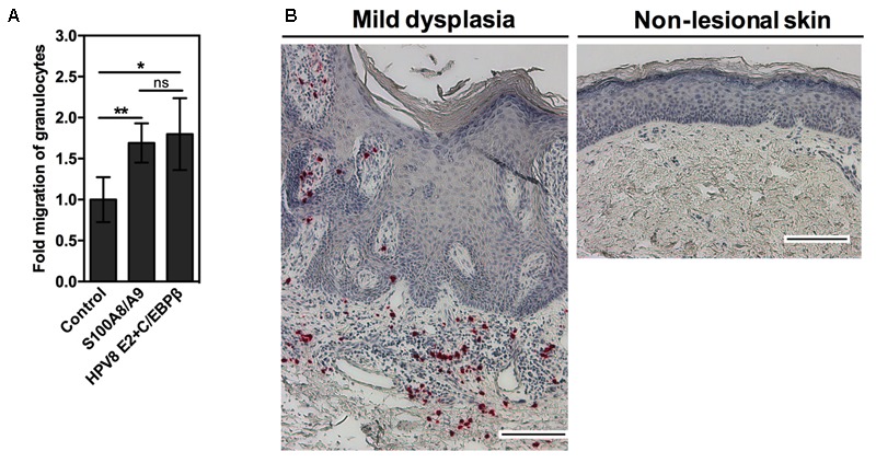
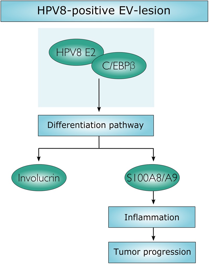
Persistent genus β-HPV (human papillomavirus) infection is a major co-factor for non-melanoma skin cancer in patients suffering from the inherited skin disease epidermodysplasia verruciformis (EV). Malignant EV lesions are particularly associated with HPV type 5 or 8. There is clinical and molecular evidence that HPV8 actively suppresses epithelial immunosurveillance by interfering with the recruitment of Langerhans cells, which may favor viral persistence. Mechanisms how persistent HPV8 infection promotes the carcinogenic process are, however, less well understood. In various tumor types chronic inflammation has a central role in tumor progression. The calprotectin complex consisting of S100A8 and S100A9 proteins has recently been identified as key driver of chronic and tumor promoting inflammation in skin carcinogenesis. It induces chemotaxis of neutrophil granulocytes and modulates inflammatory as well as immune responses. In this study, we demonstrate that skin lesions of EV-patients are massively infiltrated by inflammatory cells, including CD15+ granulocytes. At the same time we observed a very strong expression of S100A8 and S100A9 proteins in lesional keratinocytes, which was mostly confined to the suprabasal layers of the epidermis. Both proteins were hardly detected in non-lesional skin. Further experiments revealed that the HPV8 oncoproteins E6 and E7 were not involved in S100A8/A9 up-regulation. They rather suppressed differentiation-induced S100A8/A9 expression. In contrast, the viral transcription factor E2 strongly enhanced PMA-mediated S100A8/A9 up-regulation in primary human keratinocytes. Similarly, a tremendous up-regulation of both S100 proteins was observed, when minute amounts of the PMA-inducible CCAAT/enhancer binding protein β (C/EBPβ), which is expressed at low levels in the suprabasal layers of the epidermis, were co-expressed together with HPV8 E2. This confirmed our previous observation that C/EBPβ interacts and functionally synergizes with the HPV8 E2 protein in differentiation-dependent gene expression. Potent synergistic up-regulation of S100A8/A9 was seen at transcriptional and protein levels. S100A8/A9 containing supernatants from keratinocytes co-expressing HPV8 E2 and C/EBPβ significantly induced chemotaxis of granulocytes in migration assays supporting the relevance of our finding. In conclusion, our data suggest that the HPV8 E2 protein actively contributes to the recruitment of myeloid cells into EV skin lesions, which may support chronic inflammation and progression to skin cancer.

Keywords: C/EBP; E2; HPV; S100A8/A9; epidermodysplasia verruciformis; inflammation.


Figures

FIGURE 1
FIGURE 1
Strong expression of S100A8/A9 in HPV8-positive EV-lesions corresponds to an inflammatory infiltrate. (A) Serial sections of HPV8-positive EV-lesions were analyzed by IHC for S100A8 and S100A9 (magnifications in i and ii) or (B) CD45 (all red) and counterstained with hematoxylin. Scale bars 100 μm. (C) Infiltrating immune cells were counted in the stroma of mild and severe dysplasia of EV-lesions versus non-lesional skin of EV-patients based on hematoxylin counterstaining and are given as cell counts per mm2. Values represent counts ± SD from six different areas. ∗∗∗p < 0.001, unpaired t-test.
FIGURE 2
FIGURE 2
Not HPV8 E6/E7 oncoproteins but the E2 transcription factor induces both S100A8 and S100A9. S100A8/A9 mRNA levels in NFK stably expressing (A) HPV8 E6/E7 or (B) HPV8 E2 and corresponding control pLXSN cells were measured by qRT-PCR in relation to RPL13A. The amount of S100A8 and S100A9 in pLXSN control cells was set at 1. Shown are the mean values ± SD from n ≥ 2 independent experiments performed in duplicates. (C) Serial sections of OC of HPV8 E2-expressing or control pLXSN RTS3b cells were analyzed by IHC for S100A8 and S100A9 (both red) and counterstained with hematoxylin. Scale bars 100 μm. (D) NFK stably expressing HPV8 E2 or control pLXSN cells were stimulated with PMA or DMSO as a vehicle control and 24 h later S100A8 and S100A9 mRNA levels were measured by qRT-PCR in relation to RPL13A. Shown are the mean values ± SD from one representative experiment out of three performed in duplicates. ns, not significant, p < 0.05, ∗∗p < 0.01, ∗∗∗p < 0.001, unpaired t-test.
FIGURE 3
FIGURE 3
HPV8 E2 enhances PMA- or C/EBPβ-induced activation of the S100A8 promoter in human keratinocytes. (A) 0.1 × 10⁶ NFKs were seeded onto 12-well plates, next day co-transfected with 0.2 μg S100A8 reporter vector and 0.005, 0.04, or 0.2 μg HPV8 E2 expression vector and 6 h later stimulated with PMA or DMSO as a vehicle control. 0.61 × 10⁵ RTS3b cells were seeded onto 12-well plates and next day co-transfected with 0.2 μg S100A8 reporter vector together with 0.005 μg C/EBPβ and in (B) 0.005, 0.04, or 0.2 μg HPV8 E2, (C) 0.2 μg HPV8 E2 or HPV8 E2ΔC, (D) 0.005, 0.04, or 0.2 μg HPV8 E6, (E) 0.005, 0.04, or 0.2 μg HPV8 E7 or (F) HPV8 E2 and E7 expression vectors (0.2 μg each). Total amount of DNA in all transfections was adjusted up to 0.8 μg with pcDNA3.1+ empty vector. 24 h post-transfection the luciferase activity was measured and normalized to protein concentration. Control transfection was set at 1. Shown are the mean values ± SD from n ≥ 3 independent experiments performed in triplicates. ns, not significant, p < 0.05, ∗∗p < 0.01, ∗∗∗p < 0.001, ∗∗∗∗p < 0.0001, unpaired t-test.
FIGURE 4
FIGURE 4
HPV8 E2 synergizes with minute amounts of C/EBPβ to up-regulate S100A8 and S100A9 in human keratinocytes. RTS3b cells were seeded onto 6-cm dishes at a density of 0.4 × 10⁶ cells per dish and transiently co-transfected with 2.64 μg HPV8 E2 and 0.1 μg C/EBPβ expression vectors and 24 h later were analyzed for (A) S100A8 and (B) S100A9 mRNA expression by qRT-PCR in relation to RPL13A. Shown are the mean values ± SD from n = 3 independent experiments performed in duplicates. (C) For Western blot analysis 1 × 10⁶ RTS3b cells were seeded onto 10-cm dishes and next day co-transfected with 0.27 μg C/EBPβ and 7.2 μg HPV8 E2 expression vectors. Forty-eight hours later whole-cell extracts were analyzed for S100A8 (left) and S100A9 (right) protein expression. β-actin served as a loading control. Shown is one representative experiment out of n = 3. Diagrams summarize n = 3 experiments. ND, not detected; ns, not significant, ∗∗p < 0.01, ∗∗∗p < 0.001, ∗∗∗∗p < 0.0001, unpaired t-test.
FIGURE 5
FIGURE 5
Co-expression of HPV8 E2 and C/EBPβ induces migration of granulocytes in vitro. (A) Granulocyte migration toward conditioned media collected from RTS3b cells co-transfected with S100A8 and S100A9 or HPV8 E2 and C/EBPβ expression vectors or control vectors was measured by transwell chemotaxis assay. Number of migrated granulocytes toward control conditioned media was set at 1. Shown are the mean values ± SD from n = 3 independent experiments performed in duplicates. (B) Sections of EV-lesions were stained with anti-CD15 antibody by IHC (red). Counterstaining with hematoxylin reveals the segmented appearance of nuclei in CD15-positive cells indicating infiltration with granulocytes. Scale bars 100 μm. ns, not significant, p < 0.05, ∗∗p < 0.01, unpaired t-test.
FIGURE 6
FIGURE 6
Schematic presentation. Molecular mechanism, by which HPV8 E2 synergizes with C/EBPβ to induce keratinocyte differentiation and to up-regulate S100A8/A9 expression.

Similar articles

Cited by

References

    1. Agarwal C., Efimova T., Welter J. F., Crish J. F., Eckert R. L. (1999). CCAAT/enhancer-binding proteins. A role in regulation of human involucrin promoter response to phorbol ester. J. Biol. Chem. 274 6190–6194. 10.1074/jbc.274.10.6190 - DOI - PubMed
    1. Akgul B., Bostanci N., Westphal K., Nindl I., Navsaria H., Storey A., et al. (2010). Human papillomavirus 5 and 8 E6 downregulate interleukin-8 secretion in primary human keratinocytes. J. Gen. Virol. 91(Pt 4) 888–892. 10.1099/vir.0.016527-0 - DOI - PubMed
    1. Akgul B., Karle P., Adam M., Fuchs P. G., Pfister H. J. (2003). Dual role of tumor suppressor p53 in regulation of DNA replication and oncogene E6-promoter activity of epidermodysplasia verruciformis-associated human papillomavirus type 8. Virology 308 279–290. 10.1016/S0042-6822(02)00133-2 - DOI - PubMed
    1. Balasubramanian S., Agarwal C., Efimova T., Dubyak G. R., Banks E., Welter J., et al. (2000). Thapsigargin suppresses phorbol ester-dependent human involucrin promoter activity by suppressing CCAAT-enhancer-binding protein alpha (C/EBPalpha) DNA binding. Biochem. J. 350(Pt 3) 791–796. 10.1042/bj3500791 - DOI - PMC - PubMed
    1. Bresnick A. R., Weber D. J., Zimmer D. B. (2015). S100 proteins in cancer. Nat. Rev. Cancer 15 96–109. 10.1038/nrc3893 - DOI - PMC - PubMed

LinkOut - more resources