

GSK-3β-mediated regulation of cadmium-induced cell death and survival
- PMID: 29563926
- PMCID: PMC5848555
- DOI: 10.1186/s11658-018-0076-2
GSK-3β-mediated regulation of cadmium-induced cell death and survival
Abstract
Background: Previous studies indicated that cadmium (Cd) increases PI3-kinase/Akt phosphorylation, resulting in an alteration in GSK-3β activity. However, the mechanism of Cd-induced endoplasmic reticulum (ER) stress in neuronal cells has yet to be studied in needs further elucidation. We examined the role of GSK-3β in Cd-induced neuronal cell death and the related downstream signaling pathways.
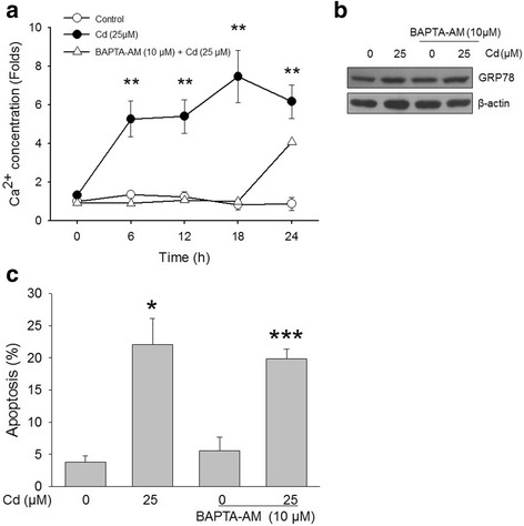
Methods: SH-SY5Y human neuroblastoma cells were treated with 10 or 20 μM BAPTA-AM and 1 μM wortmannin for 30 min and then incubated with 25 μM Cd for 12 h. Apoptotic cells were visualized via DAPI and PI staining. Data were evaluated with one-way analysis of variance (ANOVA) followed by Student's t-test. Data are expressed as the means ± SD of experiments performed at least three times.
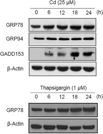
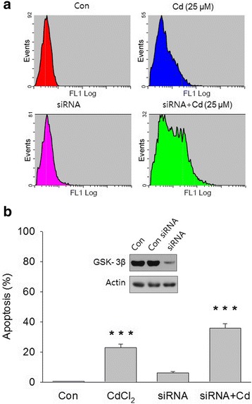
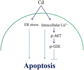
Results: Treatment of human neuronal SH-SY5Y cells with Cd induced ER, stress as evidenced by the increased expression of GRP78, which is a marker of ER stress. Cd exposure significantly increased the phosphorylation of Akt at thr308 and ser473 and that of GSK-3β at ser9 in a time-dependent manner, while the total protein levels of GSK-3β and Akt did not change. Cd-induced apoptosis was higher in GSK-3β-knockdown cells than in normal cells.
Conclusions: Our data suggest that Akt/GSK-3β signaling activated by Cd is involved in neuronal cell survival.
Keywords: Cadmium; ER-stress; GSK-3β.
Conflict of interest statement
Not applicable.Not applicable.The authors declare that they have no competing interests.Springer Nature remains neutral with regard to jurisdictional claims in published maps and institutional affiliations.
Figures

Similar articles
-
Activation of AKT1/GSK-3β/β-Catenin-TRIM11/Survivin Pathway by Novel GSK-3β Inhibitor Promotes Neuron Cell Survival: Study in Differentiated SH-SY5Y Cells in OGD Model.Mol Neurobiol. 2016 Dec;53(10):6716-6729. doi: 10.1007/s12035-015-9598-z. Epub 2015 Dec 11. Mol Neurobiol. 2016. PMID: 26660108
-
Famotidine has a neuroprotective effect on MK-801 induced toxicity via the Akt/GSK-3β/β-catenin signaling pathway in the SH-SY5Y cell line.Chem Biol Interact. 2019 Dec 1;314:108823. doi: 10.1016/j.cbi.2019.108823. Epub 2019 Sep 26. Chem Biol Interact. 2019. PMID: 31563592
-
Positive correlation between overexpression of phospho-BAD with phosphorylated Akt at serine 473 but not threonine 308 in colorectal carcinoma.Cancer Lett. 2004 Jul 16;210(2):139-50. doi: 10.1016/j.canlet.2004.01.017. Cancer Lett. 2004. PMID: 15183529
-
Valproate Attenuates Endoplasmic Reticulum Stress-Induced Apoptosis in SH-SY5Y Cells via the AKT/GSK3β Signaling Pathway.Int J Mol Sci. 2017 Feb 8;18(2):315. doi: 10.3390/ijms18020315. Int J Mol Sci. 2017. PMID: 28208696 Free PMC article.
-
Cadmium inhibits neural stem/progenitor cells proliferation via MitoROS-dependent AKT/GSK-3β/β-catenin signaling pathway.J Appl Toxicol. 2021 Dec;41(12):1998-2010. doi: 10.1002/jat.4179. Epub 2021 May 11. J Appl Toxicol. 2021. PMID: 33977565 Review.
Cited by
-
Activation of Pgk1 Results in Reduced Protein Aggregation in Diverse Neurodegenerative Conditions.Mol Neurobiol. 2023 Sep;60(9):5090-5101. doi: 10.1007/s12035-023-03389-6. Epub 2023 May 30. Mol Neurobiol. 2023. PMID: 37249790
-
The Effects of Seleno-Methionine in Cadmium-Challenged Human Primary Chondrocytes.Pharmaceuticals (Basel). 2024 Jul 12;17(7):936. doi: 10.3390/ph17070936. Pharmaceuticals (Basel). 2024. PMID: 39065786 Free PMC article.
-
Cadmium exposure alters steroid receptors and proinflammatory cytokine levels in endothelial cells in vitro: a potential mechanism of endocrine disruptor atherogenic effect.J Endocrinol Invest. 2019 Jun;42(6):727-739. doi: 10.1007/s40618-018-0982-1. Epub 2018 Nov 26. J Endocrinol Invest. 2019. PMID: 30478740
-
Mechanisms of Cadmium Neurotoxicity.Int J Mol Sci. 2023 Nov 21;24(23):16558. doi: 10.3390/ijms242316558. Int J Mol Sci. 2023. PMID: 38068881 Free PMC article. Review.
-
MicroRNA-429 inhibits neuroblastoma cell proliferation, migration and invasion via the NF-κB pathway.Cell Mol Biol Lett. 2020 Feb 13;25:5. doi: 10.1186/s11658-020-0202-9. eCollection 2020. Cell Mol Biol Lett. 2020. PMID: 32082390 Free PMC article.
References
-
- Kwon OY, Kim YJ, Choi Y, Kim H, Song C, Shong M. The endoplasmic reticulum chaperone GRP94 is induced in the thyrocytes by cadmium. Z Naturforsch C. 1999;54(7–8):573–577. - PubMed
MeSH terms
Substances
LinkOut - more resources
Full Text Sources
Other Literature Sources
Molecular Biology Databases
Research Materials
Miscellaneous
