

Giant Peripheral Ossifying Fibroma of the Posterior Mandible-A Rare Case Report
- PMID: 29563937
- PMCID: PMC5844686
Giant Peripheral Ossifying Fibroma of the Posterior Mandible-A Rare Case Report
Abstract
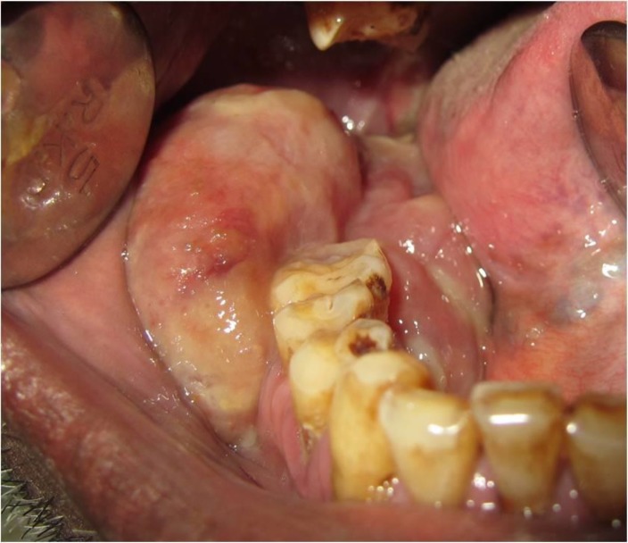
Large, atypical peripheral ossifying fibromas are known as giant peripheral ossifying fibromas. These lesions have often been associated with heterogeneous clinical and radiographic characteristics subsequently leading to their misdiagnosis. Biopsies have been the gold standard for the diagnosis of such lesions. This study reports on an acute presentation of giant peripheral ossifying fibroma, clinically mimicking a malignant lesion due to its atypical presentation along with its characteristic histological features, which led to the establishment of the diagnosis.
Keywords: Giant; gingival growths; gingival neoplasms; ossifying fibroma.
Conflict of interest statement
The authors have no financial relationships with any organization.
Figures


Similar articles
-
Giant peripheral ossifying fibroma with coincidental squamous cell carcinoma: a case report.J Med Case Rep. 2021 Dec 19;15(1):599. doi: 10.1186/s13256-021-03187-5. J Med Case Rep. 2021. PMID: 34922609 Free PMC article.
-
A giant peripheral ossifying fibroma of the mandible: A rare case report.Int J Surg Case Rep. 2024 Jan;114:109161. doi: 10.1016/j.ijscr.2023.109161. Epub 2023 Dec 20. Int J Surg Case Rep. 2024. PMID: 38157625 Free PMC article.
-
Peripheral ossifying fibroma: A case report.J Indian Soc Periodontol. 2013 Nov;17(6):819-22. doi: 10.4103/0972-124X.124533. J Indian Soc Periodontol. 2013. PMID: 24554899 Free PMC article.
-
Giant peripheral ossifying fibroma: a case report and clinicopathologic review of 10 cases from the literature.Head Neck Pathol. 2013 Dec;7(4):356-60. doi: 10.1007/s12105-013-0452-1. Epub 2013 Jul 16. Head Neck Pathol. 2013. PMID: 23857548 Free PMC article. Review.
-
Peripheral ossifying fibroma. Report of a case and review of the literature.Med Oral. 2001 Mar-Apr;6(2):135-41. Med Oral. 2001. PMID: 11500629 Review. English, Spanish.
Cited by
-
Surgical resection of a giant peripheral ossifying fibroma in mouth floor managed with fiberscopic intubation.Clin Case Rep. 2020 Nov 6;9(1):180-184. doi: 10.1002/ccr3.3494. eCollection 2021 Jan. Clin Case Rep. 2020. PMID: 33489156 Free PMC article.
-
Giant peripheral ossifying fibroma with coincidental squamous cell carcinoma: a case report.J Med Case Rep. 2021 Dec 19;15(1):599. doi: 10.1186/s13256-021-03187-5. J Med Case Rep. 2021. PMID: 34922609 Free PMC article.
-
Giant Ossifying Fibroma (Epulis) in an Elderly Patient-A Case Report.Indian J Otolaryngol Head Neck Surg. 2025 Feb;77(2):1050-1054. doi: 10.1007/s12070-024-05244-z. Epub 2024 Dec 6. Indian J Otolaryngol Head Neck Surg. 2025. PMID: 40070767
References
-
- Rajendran R, Sivapathasundaram B, editors. Shafer’s Textbook of Oral Pathology. 7thed. New Delhi: Elsevier; 2012. pp. 133–134.
-
- Neville BW, Damm DD, Allen CM, Buoquot JE. Oral and Maxillofacial Pathology. 2nd ed. Philadelphia: WB Saunders; pp. 451–452.
-
- Gardner DG. Peripheral odontogenic fibroma: An attempt of classifcation. Oral Surg Oral Med Oral Pathol. 1982;54(1):40–8. - PubMed
-
- Silva CO, Sallum AW, do Couto-Filho CE, Costa Pereira AA, Hanemann JA, Tatakis DN. Localized gingival enlargement associated with alveolar process expansion: peripheral ossifying fibroma coincident with central odontogenic fibroma. J Periodontol. 2007;78(7):1354–9. - PubMed
Publication types
LinkOut - more resources
Full Text Sources