

Multicentric Glioblastoma Multiforme Mimicking Optic Neuritis
- PMID: 29563957
- PMCID: PMC5858864
- DOI: 10.1080/01658107.2017.1350194
Multicentric Glioblastoma Multiforme Mimicking Optic Neuritis
Abstract
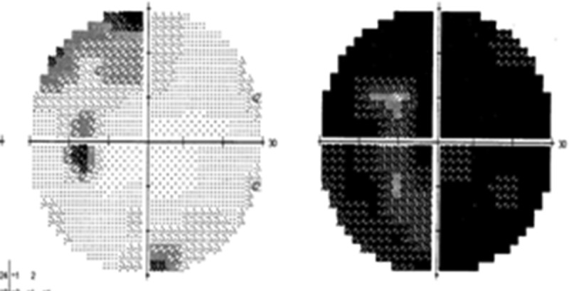
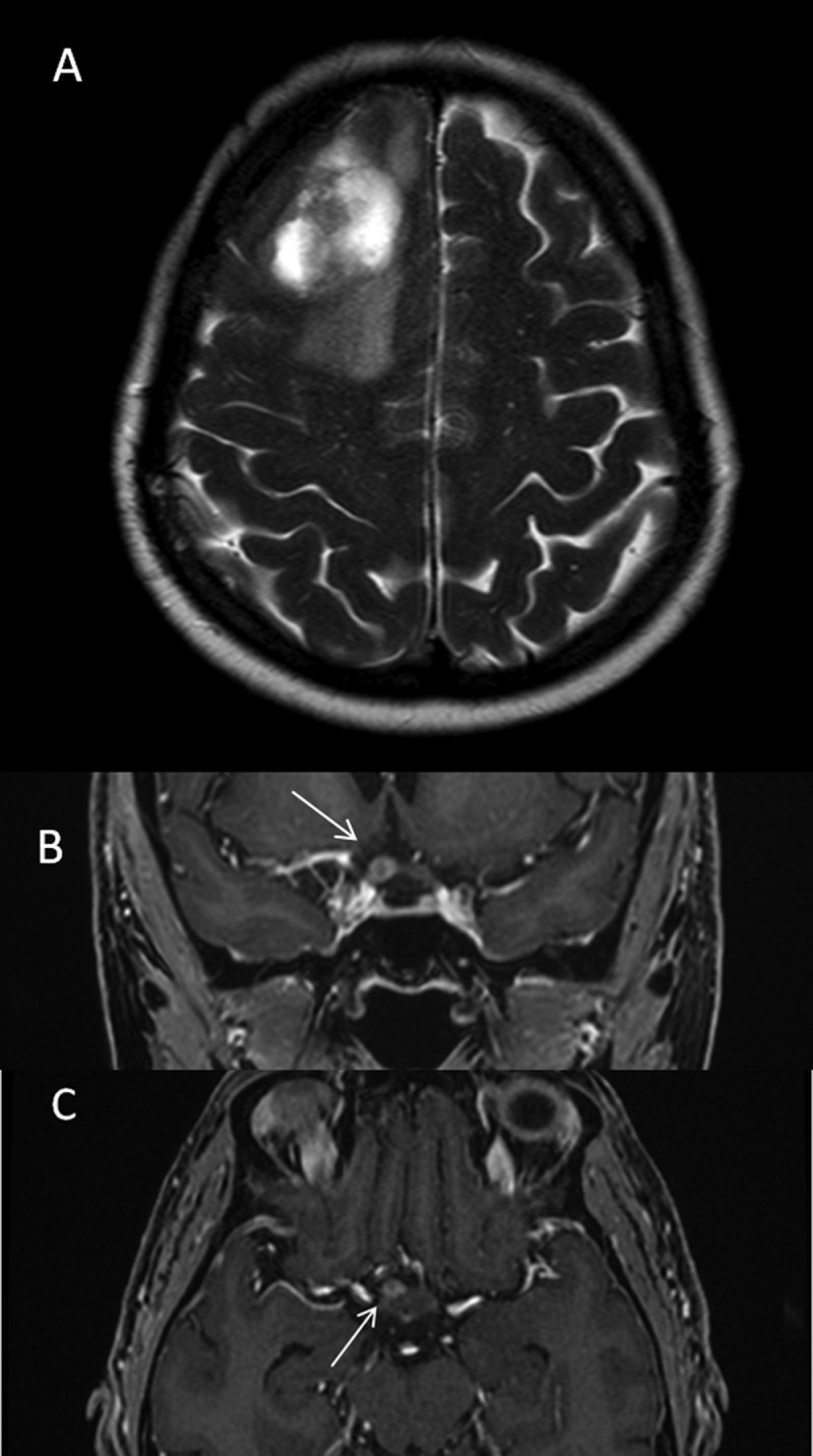
A 49-year-old previously healthy woman presented with acute painless visual loss in the right eye, a right relative afferent pupillary defect, and a normal fundus examination. She was diagnosed with retrobulbar "optic neuritis" and given a course of intravenous steroids. Despite treatment, however, she continued to lose vision and serial visual field testing confirmed a junctional scotoma in the fellow eye. Cranial magnetic resonance imaging (MRI) showed a mass at the junction between the right optic nerve and the anterior aspect of the chiasm and a right frontal lesion that proved to be multicentric glioblastoma multiforme. Clinicians should be aware of the possibility of aggressive neoplasm in the differential diagnosis of retrobulbar optic neuritis.
Keywords: Glioblastoma multiforme; malignant optic nerve glioma of the adult; optic nerve tumour; optic neuritis.
Figures

Similar articles
-
Multifocal malignant optic glioma of adulthood presenting as acute anterior optic neuropathy.J Clin Neurosci. 2011 Jul;18(7):974-7. doi: 10.1016/j.jocn.2010.12.010. Epub 2011 May 6. J Clin Neurosci. 2011. PMID: 21550255
-
Glioblastoma multiforme mimicking optic neuritis.Am J Ophthalmol Case Rep. 2020 Jan 7;17:100594. doi: 10.1016/j.ajoc.2020.100594. eCollection 2020 Mar. Am J Ophthalmol Case Rep. 2020. PMID: 32395666 Free PMC article.
-
At this junction….Surv Ophthalmol. 2022 Nov-Dec;67(6):1711-1716. doi: 10.1016/j.survophthal.2021.08.001. Epub 2021 Aug 6. Surv Ophthalmol. 2022. PMID: 34364902
-
Unilateral adult malignant optic nerve glioma.Graefes Arch Clin Exp Ophthalmol. 2004 Sep;242(9):741-8. doi: 10.1007/s00417-004-0905-z. Graefes Arch Clin Exp Ophthalmol. 2004. PMID: 15085353 Review.
-
The natural history of optic neuritis.Rev Neurol Dis. 2006 Spring;3(2):45-56. Rev Neurol Dis. 2006. PMID: 16819420 Review.
Cited by
-
Long noncoding RNA LINC00511 induced by SP1 accelerates the glioma progression through targeting miR-124-3p/CCND2 axis.J Cell Mol Med. 2019 Jun;23(6):4386-4394. doi: 10.1111/jcmm.14331. Epub 2019 Apr 11. J Cell Mol Med. 2019. PMID: 30973678 Free PMC article.
-
A Novel Predictive Model Utilizing Retinal Microstructural Features for Estimating Survival Outcome in Patients with Glioblastoma.Res Sq [Preprint]. 2024 May 17:rs.3.rs-4420925. doi: 10.21203/rs.3.rs-4420925/v1. Res Sq. 2024. Update in: Clin Neurol Neurosurg. 2025 Mar;250:108790. doi: 10.1016/j.clineuro.2025.108790. PMID: 38798600 Free PMC article. Updated. Preprint.
-
Multicentric Glioma: An Ideal Model to Reveal the Mechanism of Glioma.Front Oncol. 2022 Jun 7;12:798018. doi: 10.3389/fonc.2022.798018. eCollection 2022. Front Oncol. 2022. PMID: 35747806 Free PMC article. Review.
-
The varieties of junctional scotoma: 17 cases, a review, and a taxonomy.Eye (Lond). 2025 Jun;39(9):1673-1687. doi: 10.1038/s41433-025-03789-z. Epub 2025 Apr 22. Eye (Lond). 2025. PMID: 40263597 Review.
-
A novel predictive model utilizing retinal microstructural features for estimating survival outcome in patients with glioblastoma.Clin Neurol Neurosurg. 2025 Mar;250:108790. doi: 10.1016/j.clineuro.2025.108790. Epub 2025 Feb 17. Clin Neurol Neurosurg. 2025. PMID: 39987704
References
-
- Wilbrand H, Saenger A.. The neurology of the eyes. J Bergmann 1904;3:98–120.
-
- Wilbrand H. Schema des Verlaufs der Sehnervenfasern durch das Chiasma. Z Aufenheilkd 1926;59:135–144.
-
- Shin RK, Qureshi RA, Harris NR, Bakar D, Li TP, Jafri S, Tang CM.. Wilbrand knee. Neurology 2014;82:459–460. - PubMed
Publication types
LinkOut - more resources
Full Text Sources
Other Literature Sources