

Case Reports
doi: 10.1080/01658107.2017.1355395.
eCollection 2018 Apr.
Papilloedema and Autoimmune Retinopathy from Systemic Lupus Erythematosus
Affiliations
- PMID: 29563958
- PMCID: PMC5858861
- DOI: 10.1080/01658107.2017.1355395
Item in Clipboard
Case Reports
Papilloedema and Autoimmune Retinopathy from Systemic Lupus Erythematosus
Neuroophthalmology.
.
Abstract
A 33-year-old female presented with bilateral papilloedema and constricted visual fields from autoimmune retinopathy. She then developed a painful peripheral neuropathy that led to further work-up and the diagnosis of systemic lupus erythematosus. Papilloedema and autoimmune retinopathy from systemic lupus erythematosus is a unique presentation.
Keywords: Autoimmune retinopathy; idiopathic intracranial hypertension; papilloedema; pseudotumor cerebri; systemic lupus erythematosus.
Figures
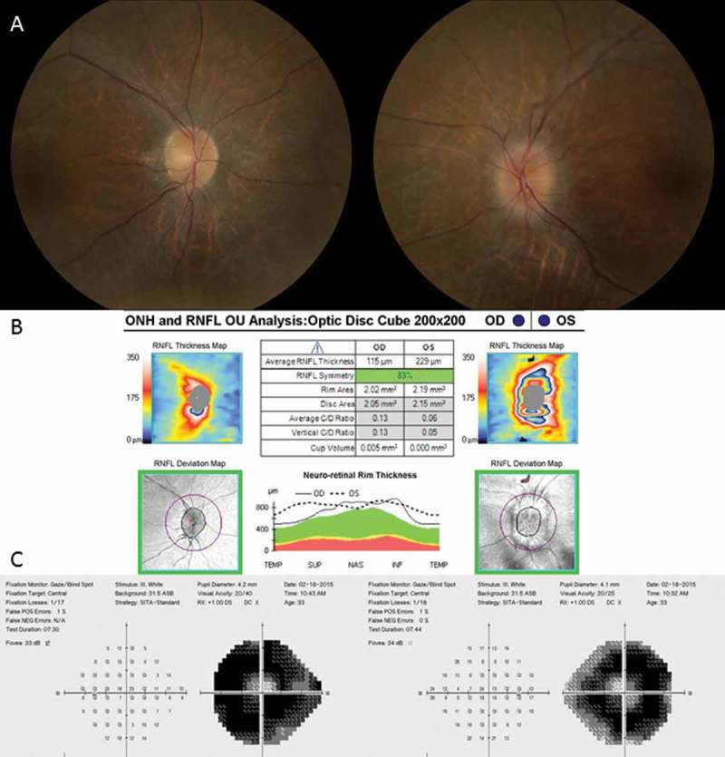
Fundus photos demonstrate bilateral disc oedema, left greater than right (A). Optical coherence tomography (OCT) shows thickening of the retinal nerve fibre layer, left greater than right (B). 24-2 Humphrey visual fields show severe constriction of both eyes (C).

Manual visual fields demonstrate bilateral ring scotomas (A), which correspond to areas of ellipsoid photoreceptor loss in the parafoveal region on OCT (B). ERG shows bilateral severe reduction of both rod and cone function (C).

Five-month follow-up later demonstrates significant improvement in the disc oedema (A). Humphrey visual fields show severe constriction in both eyes, which is unchanged (B).
References
-
- Kuyucu S, Argin A, Kuyucu N, Ozen S. Systemic lupus erythematosus presenting with pseudotumor cerebri: a rare association. Turk J Pediatr 2007;49:98–101. - PubMed
-
- Parnass SM, Goodwin JA, Patel DV, Levinson DJ, Reinhard JD. Dural sinus thrombosis: a mechanism for pseudotumor cerebri in systemic lupus erythematosus. J Rheumatol 1987;14:152–155. - PubMed
Publication types
LinkOut - more resources
Full Text Sources
Other Literature Sources