. 2018 Apr;15(4):3836-3846.
doi: 10.3892/etm.2018.5893. Epub 2018 Feb 26.

Protective effects of paeonol on subacute/chronic brain injury during cerebral ischemia in rats

Affiliations

Protective effects of paeonol on subacute/chronic brain injury during cerebral ischemia in rats

Bing Zhao et al. Exp Ther Med. 2018 Apr.

Abstract

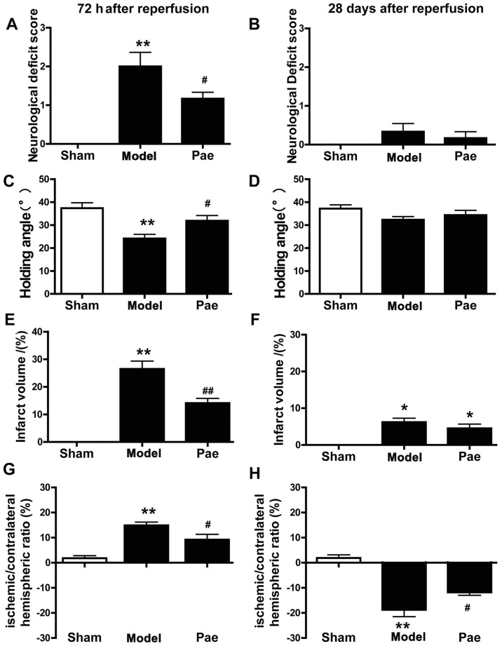
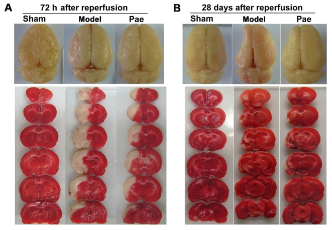
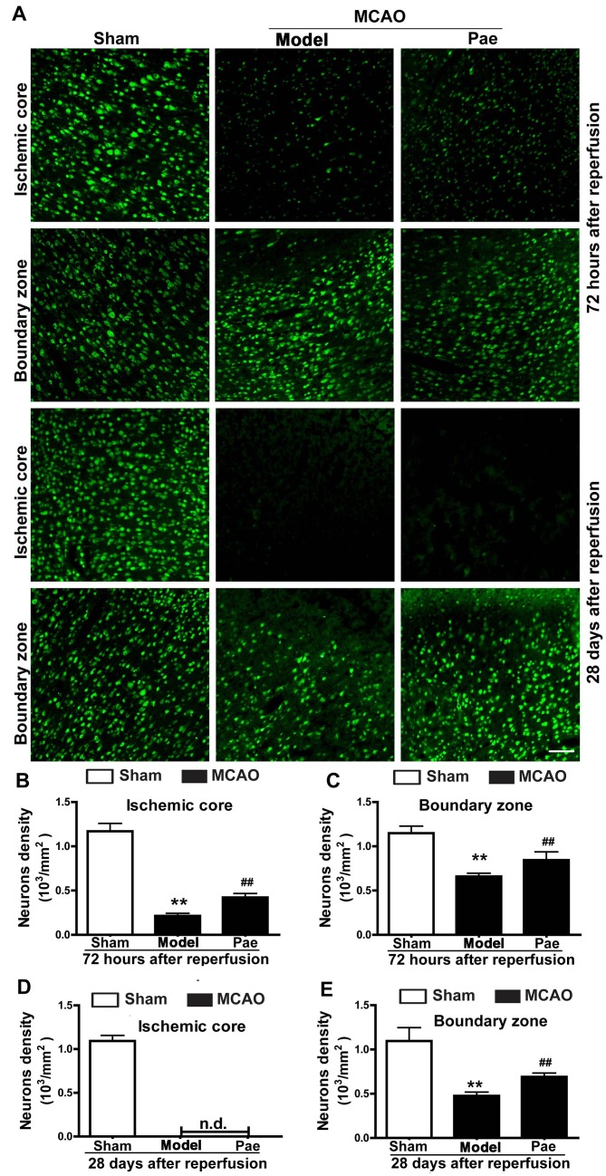
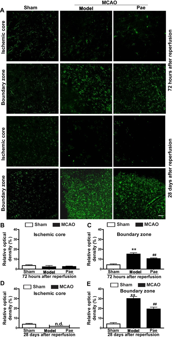
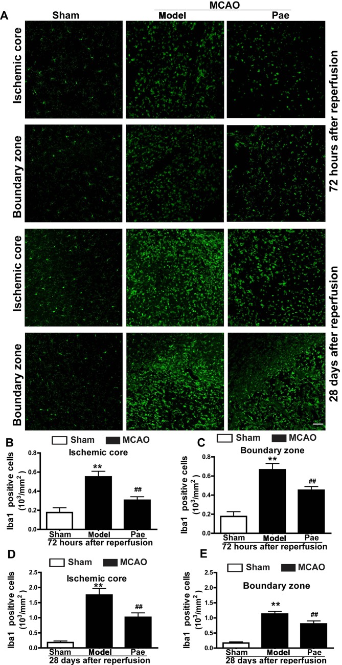
Ischemic stroke is a highly complex pathological process that is divided into acute, subacute and chronic phases. Paeonol is a biologically active natural product with a variety of pharmacological effects, including those on neuronal activity. However, the effects of paeonol on subacute/chronic ischemic stroke have remained to be elucidated. The present study was designed to investigate the effects of paeonol against subacute and chronic cerebral ischemic injury and to explore the possible underlying mechanisms. Male adult Sprague Dawley rats were randomly divided into a sham group (treated with saline), a model group [subjected to middle cerebral artery occlusion (MCAO) and treated with saline] and a paeonol-treated group (MCAO + paeonol at 25 mg/kg). Behavioral impairment, infarct volume and ischemic/contralateral hemispheric ratios were assessed at 72 h and at 28 days after MCAO, respectively. Immunofluorescence was employed to determine the neuronal damage and glial responses after MCAO. Compared with the model group, paeonol treatment significantly attenuated behavioral impairment, ischemic infarct volume and moderate cerebral edema in the ischemic brain at 72 h, as well as brain atrophy at 28 days after reperfusion. Furthermore, paeonol treatment ameliorated neuronal damage in the ischemic core and boundary zone regions at 72 h after reperfusion and in the boundary zone at 28 days after reperfusion. In addition, paeonol treatment reduced the proliferation of astrocytes in the boundary zone, and inhibited microglial activation in the ischemic core and boundary zone regions at 72 h and 28 days after reperfusion. These results demonstrated the protective effects of paeonol against subacute/chronic cerebral ischemia, and the mechanism of action may include subacute/chronic microglial activation and astrocyte proliferation.

Keywords: cerebral ischemia; paeonol; rat; subacute/chronic brain injury.


Figures

Figure 1.
Figure 1.
Effects of paeonol on neurological dysfunction, infarct volume and the ischemic/contralateral hemispheric ratio after focal cerebral ischemia. (A-D) Paeonol treatment significantly ameliorated the neurological deficit score and holding angle at 72 h after reperfusion compared with those in the model group; however, it did not affect the already recovered neurological deficit score and holding angle at 28 days after reperfusion. (E and F) Paeonol treatment significantly decreased the infarct volume at 72 h after reperfusion compared with that in the model group; however, it did not affect the already decreased infarct volume at 28 days after reperfusion. (G and H) The ischemic/contralateral hemispheric ratio was increased (brain edema) at 72 h and decreased (brain atrophy) at 28 days after reperfusion, and these changes were significantly ameliorated by paeonol treatment. Values are expressed as the mean ± standard error of the mean from eight rats per group. *P<0.05 and **P<0.01 compared with sham group, #P<0.05 and ##P<0.01 compared with model group. Pae, paeonol.
Figure 2.
Figure 2.
Effects of paeonol on ischemic brain injury. Representative images of total brains indicated brain lesions at (A) 72 h and (B) 28 days after reperfusion (upper panel). Representative images of TTC-stained coronal slices with 2 mm-thickness indicated ischemic lesions at (A) 72 h and (B) 28 days after reperfusion (lower panel). After TTC staining, the viable tissue stained deep red, whereas the infarct area was white in color. TTC, 2,3,5-triphenyltetrazolium chloride; Pae, paeonol.
Figure 3.
Figure 3.
Effects of paeonol on neuronal injury at 72 h and 28 days after reperfusion in rats. (A) Representative photomicrographs indicated neurons by immunostaining for NeuN at 72 h and at 28 days after reperfusion (scale bar, 100 µm) and (B-E) quantification results. In the ischemic core, the reduction of neuronal density was significantly attenuated by paeonol treatment at 72 h after reperfusion, whereas NeuN-expressing cells completely disappeared at 28 days after reperfusion. In the boundary zone, the decrease of neuronal density was significantly attenuated by paeonol treatment at 72 h and 28 days after reperfusion. Values are expressed as the mean ± standard error of the mean from eight rats per group. **P<0.01 compared with sham group, ##P<0.01 compared with model group. Pae, paeonol; MCAO, middle cerebral artery occlusion; n.d., not detectable; NeuN, neuronal nuclei.
Figure 4.
Figure 4.
Effects of paeonol on astrocyte proliferation at 72 h and 28 days after reperfusion in rats. (A) Representative photomicrographs indicating glial fibrillary acidic protein-immunopositive astrocytes at 72 h and 28 days after reperfusion (scale bar, 100 µm) and (B-E) quantification results. At 72 h after reperfusion, astrocytes were initially increased in the boundary zone, and paeonol treatment significantly suppressed the changes in the boundary zone, but not those in the ischemic core. At 28 days after reperfusion, astrocytes were significantly increased in the boundary zone, and paeonol treatment significantly attenuated the astrogliosis in the boundary zone, but not that in the ischemic core. Values are expressed as the mean ± standard error of the mean from eight rats per group. **P<0.01 compared with sham group, ##P<0.01 compared with model group. Pae, paeonol; MCAO, middle cerebral artery occlusion; n.d., not detectable.
Figure 5.
Figure 5.
Effects of paeonol on microglial activation at 72 h and 28 days after reperfusion in rats. (A) Representative photomicrographs displaying Iba-1-immunopositive microglia in the ischemic core and boundary zone regions at 72 h and 28 days after reperfusion (scale bar, 100 µm) and (B-E) quantification results. At 72 h after reperfusion, Iba-1-positive microglia initially increased in the ischemic core and the boundary zone regions, and these changes were significantly inhibited by paeonol treatment. At 28 days after reperfusion, microglial cells in the ischemic core and boundary zone regions were significantly increased, which was attenuated by paeonol treatment. Values are expressed as the mean ± standard error of the mean from eight rats per group. **P<0.01 compared with sham group, ##P<0.01 compared with model group. Pae, paeonol; MCAO, middle cerebral artery occlusion; Iba-1, ionized calcium binding adaptor molecule 1.

Similar articles

Cited by

References

    1. Hankey GJ. Stroke. Lancet. 2017;389:641–654. doi: 10.1016/S0140-6736(16)30962-X. - DOI - PubMed
    1. Jauch EC, Saver JL, Adams HP, Jr, Bruno A, Connors JJ, Demaerschalk BM, Khatri P, McMullan PW, Jr, Qureshi AI, Rosenfield K, et al. Guidelines for the early management of patients with acute ischemic stroke: A guideline for healthcare professionals from the American Heart Association/American Stroke Association. Stroke. 2013;44:870–947. doi: 10.1161/STR.0b013e318284056a. - DOI - PubMed
    1. Li L, Stary CM. Targeting glial mitochondrial function for protection from cerebral ischemia: Relevance, mechanisms, and the role of MicroRNAs. Oxid Med Cell Longev. 2016;2016:6032306. doi: 10.1155/2016/6032306. - DOI - PMC - PubMed
    1. Hacke W, Kaste M, Bluhmki E, Brozman M, Dávalos A, Guidetti D, Larrue V, Lees KR, Medeghri Z, Machnig T, et al. Thrombolysis with alteplase 3 to 4.5 h after acute ischemic stroke. N Engl J Med. 2008;359:1317–1329. doi: 10.1056/NEJMoa0804656. - DOI - PubMed
    1. Shichita T, Ago T, Kamouchi M, Kitazono T, Yoshimura A, Ooboshi H. Novel therapeutic strategies targeting innate immune responses and early inflammation after stroke. J Neurochem. 2012;123(Suppl 2):S29–S38. doi: 10.1111/j.1471-4159.2012.07941.x. - DOI - PubMed