. 2018 Apr;15(4):3892-3898.
doi: 10.3892/etm.2018.5899. Epub 2018 Feb 27.

Correlation between the expression of Drp1 in vascular endothelial cells and inflammatory factors in hypertension rats

Affiliations

Correlation between the expression of Drp1 in vascular endothelial cells and inflammatory factors in hypertension rats

Xinghui Liu et al. Exp Ther Med. 2018 Apr.

Abstract

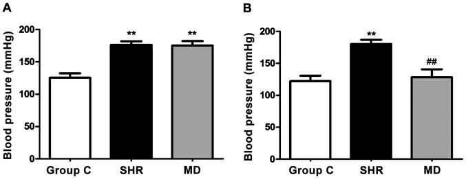
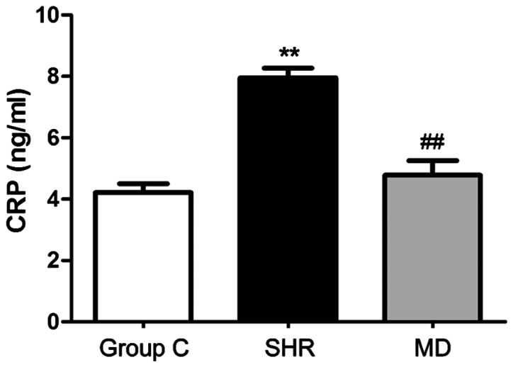
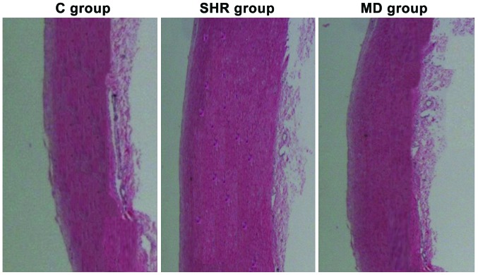
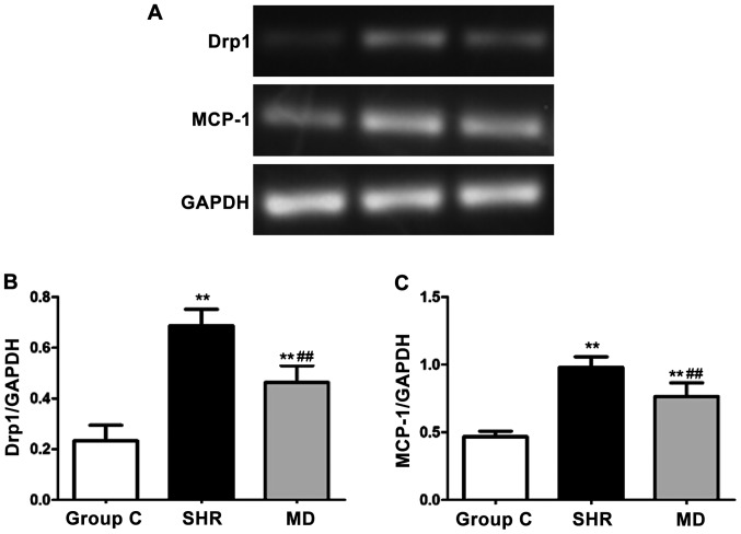
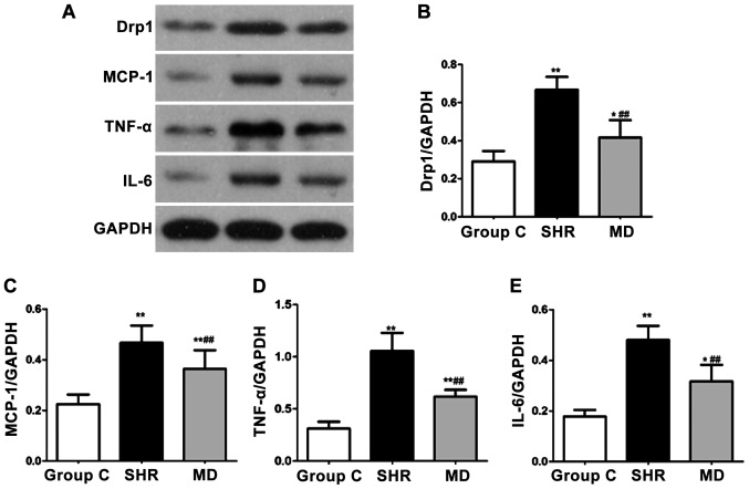
The objective of this study was to investigate the expression level of dynamin-related protein 1 (Drp1) in vascular endothelium of hypertension rats and its correlation with expression of inflammatory factors. Twenty spontaneous hypertension rats (SHR) were randomly divided into SHR group (n=10) and inhibition group (MD group, n=10), and the Sprague Dawley rats were enrolled as the control group (C group, n=10). For rats in the MD group, Mdivi-1, a mitochondrial division inhibitor, was given in dosage of 25 mg/kg. After 4 weeks of administration, blood pressure was measured via tail-artery blood pressure measurement. The blood samples collected from the abdominal aorta of rats were used to assay the C-reaction protein (CRP) concentration in serum through radioimmunoassay. Hematoxylin and eosin (H&E) staining was performed for sections of thoracic aorta for morphological observation and measurement of medial thickness. Enzyme-linked immunosorbant assay (ELISA), semi-quantitative real-time polymerase chain reaction (RT-PCR) and western blotting was carried out for detecting the expression levels of interleukin-6 (IL-6) and tumor necrosis factor-α (TNF-α). Drp1 and monocyte chemotactic protein 1 (MCP-1). After 4 weeks of drug administration, the blood pressure in the MD group was significantly higher (P<0.01). The medial thickness of the thoracic aorta in the MD group was significantly decreased in comparison with the SHR group (P<0.01). The results of ELISA showed that compared with the SHR group, the expression levels of IL-6 and TNF-α in the MD group were remarkably decreased (P<0.01). Semi-quantitative RT-PCR results indicated that the mRNA expression levels of Drp1 and MCP-1 in the MD group were significantly lower than those in the SHR group (P<0.05). In the SHR rats, after administration of Mdivi-1, the expression of Drp1 is decreased, which contributes to the alleviation in inflammatory reactions and protects the vessels in SHR rats.

Keywords: dynamin-related protein 1; inflammatory factors; spontaneous hypertension rat; vessels.


Figures

Figure 1.
Figure 1.
Variation in blood pressure of rats in each group. (A) Blood pressure of rats in each group before drug administration. The blood pressure of rats in the spontaneous hypertension rat (SHR) group and the MD group is significantly higher than that in the C group, and the difference has statistical significance (**P<0.01). (B) After 4 weeks of drug administration, variations in blood pressure of rats in each group. The blood pressure of rats in the MD group is significantly lower than that in the SHR group (##P<0.01).
Figure 2.
Figure 2.
Variation in C-reaction protein (CRP) levels in serum of rats in each group: The concentration of CRP in serum of rats in the C group is significantly lower than those in the spontaneous hypertension rat (SHR) group and the MD group (**P<0.01); the level of CRP in serum of rats in the MD group is lower than that in the SHR group, and the difference has statistical significance (##P<0.01).
Figure 3.
Figure 3.
Detecting the variation in media of thoracic aorta of rats in each group using hematoxylin and eosin (H&E) staining. Compared with the C group, the media of thoracic aorta of rats in the spontaneous hypertension rat (SHR) group are significantly thickened; while the media in the MD group are attenuated.
Figure 4.
Figure 4.
Detection of the expression levels of inflammatory factors via ELISA. (A) The level of IL-6. (B) The level of TNF-α. Compared with the C group, the levels of IL-6 and TNF-α of rats in the SHR group and the MD group are significantly elevated, and the differences have statistical significance (**P<0.01, *P<0.05); the levels of IL-6 and TNF-α in serum of rats in the MD group are lower than those in the MD group (##P<0.01).
Figure 5.
Figure 5.
Detection of expression levels of Drp1 and MCP-1 via semi-quantitative PCR. Compared with the C group, the mRNA expression levels of Drp1 and MCP-1 of rats in the SHR group and the MD group are significantly elevated, and the differences have statistical significance (**P<0.01); in comparison with the SHR group, the mRNA expression levels of Drp1 and MCP-1 of rats in the MD group are decreased (##P<0.01).
Figure 6.
Figure 6.
Detection of expression levels of proteins via western blotting assay. Compared with the C group, the protein expression levels of Drp1, MCP-1, IL-6 and TNF-α of rats in the spontaneous hypertension rat (SHR) group and the MD group are significantly increased (*P<0.05, **P<0.01); in comparison with the SHR group, significant decreases are identified in the protein expression levels of Drp1, MCP-1, IL-6 and TNF-α of rats in the MD group, and the differences have statistical significance (##P<0.01).

Similar articles

Cited by

References

    1. Song CL, Zhang X, Liu YK, Yue WW, Wu H. Heart rate turbulence in masked hypertension and white-coat hypertension. Eur Rev Med Pharmacol Sci. 2015;19:1457–1460. - PubMed
    1. Misárková E, Behuliak M, Bencze M, Zicha J. Excitation-contraction coupling and excitation-transcription coupling in blood vessels: Their possible interactions in hypertensive vascular remodeling. Physiol Res. 2016;65:173–191. - PubMed
    1. Kai H, Kudo H, Takayama N, Yasuoka S, Aoki Y, Imaizumi T. Molecular mechanism of aggravation of hypertensive organ damages by short-term blood pressure variability. Curr Hypertens Rev. 2014;10:125–133. doi: 10.2174/1573402111666141217112655. - DOI - PMC - PubMed
    1. Rubattu S, Stanzione R, Volpe M. Mitochondrial dysfunction contributes to hypertensive target organ damage: lessons from an animal model of human disease. Oxid Med Cell Longev. 2016;2016:1067801. doi: 10.1155/2016/1067801. - DOI - PMC - PubMed
    1. Zhang J, Fallahzadeh MK, McCullough PA. Aging male spontaneously hypertensive rat as an animal model for the evaluation of the interplay between contrast-induced acute kidney injury and cardiorenal syndrome in humans. Cardiorenal Med. 2016;7:1–10. doi: 10.1159/000447542. - DOI - PMC - PubMed