Fungal keratitis caused by rare organisms
- PMID: 29564417
- PMCID: PMC5859421
- DOI: 10.1016/j.joco.2017.08.004
Fungal keratitis caused by rare organisms
Abstract
Purpose: To report two rare cases of filamentous fungal keratitis.
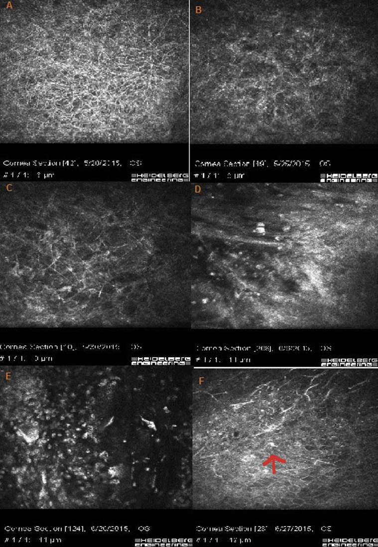
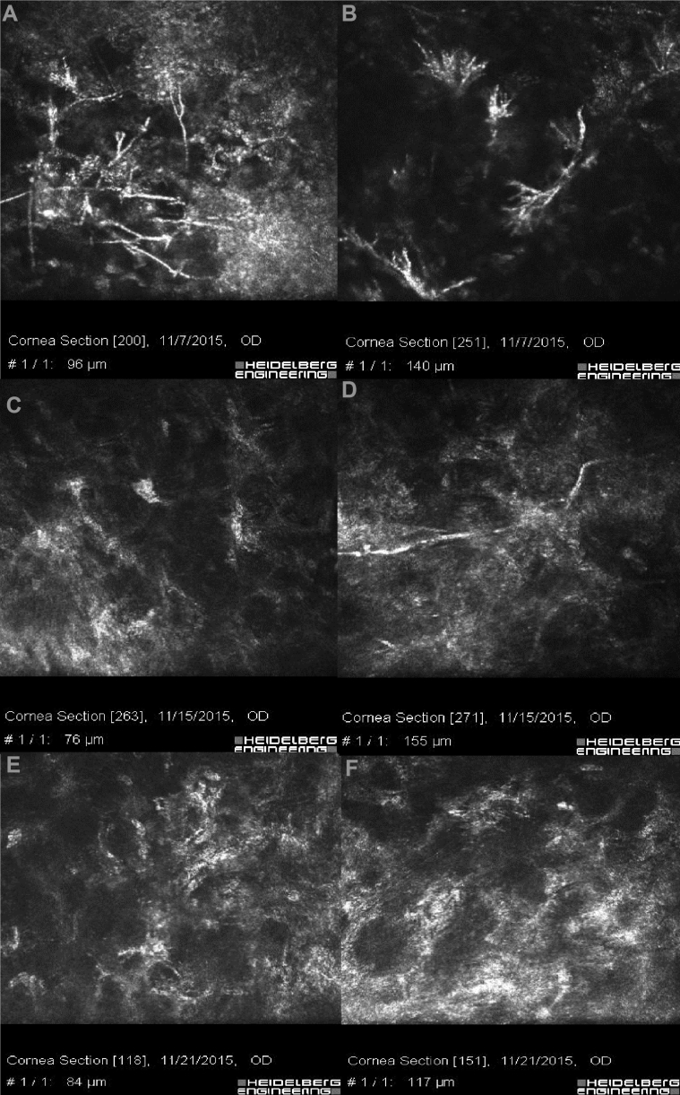
Methods: Two non-consecutive patients presented with suspicious fungal keratitis. After performing the smear and culture, medical therapy was started for them. They underwent slit photography and in vivo confocal microscopy (IVCM) in their follow-up visits.
Results: The patients were 33-year-old and 56-year-old farmer men. They both mentioned a history of ocular trauma by plants. During their follow-up visits, corneal infiltration density and fungal hyphae density decreased in slit-lamp biomicroscopy and IVCM, respectively. The corresponding organisms were Pseudallescheria boydii (P. boydii) and Colletotrichum coccodes.
Conclusions: It is important to be aware of these rare organisms and their antibiotic susceptibility. There was not any specific confocal feature for the presented fungal keratitis that was different from other filamentous fungal hyphae; however, confocal scan is a good choice to follow the response to the treatment.
Keywords: Colletotrichum coccodes; Fungal keratitis; Pseudallescheria boydii.
Figures


Similar articles
-
Exophiala phaeomuriformis Fungal Keratitis: Case Report and In Vivo Confocal Microscopy Findings.Eye Contact Lens. 2017 Mar;43(2):e4-e6. doi: 10.1097/ICL.0000000000000193. Eye Contact Lens. 2017. PMID: 26513718
-
Fungal keratitis caused by Pseudallescheria boydii: clinical and mycological characteristics.J Ophthalmic Inflamm Infect. 2021 Sep 24;11(1):30. doi: 10.1186/s12348-021-00255-1. J Ophthalmic Inflamm Infect. 2021. PMID: 34557976 Free PMC article.
-
Application of image recognition-based automatic hyphae detection in fungal keratitis.Australas Phys Eng Sci Med. 2018 Mar;41(1):95-103. doi: 10.1007/s13246-017-0613-8. Epub 2017 Dec 28. Australas Phys Eng Sci Med. 2018. PMID: 29285615
-
Infectious Keratitis Caused by Rare and Emerging Micro-Organisms.Curr Eye Res. 2020 Jul;45(7):761-773. doi: 10.1080/02713683.2019.1708407. Epub 2020 Jan 8. Curr Eye Res. 2020. PMID: 31870176 Review.
-
Clinical and mycological characteristics of keratitis caused by Colletotrichum gloeosporioides: A case report and review of literature.J Infect Dev Ctries. 2021 Mar 7;15(2):301-305. doi: 10.3855/jidc.14492. J Infect Dev Ctries. 2021. PMID: 33690215 Review.
Cited by
-
Scedosporiosis and lomentosporiosis: modern perspectives on these difficult-to-treat rare mold infections.Clin Microbiol Rev. 2024 Jun 13;37(2):e0000423. doi: 10.1128/cmr.00004-23. Epub 2024 Mar 29. Clin Microbiol Rev. 2024. PMID: 38551323 Free PMC article. Review.
-
Fungal keratitis in Iran: Risk factors, clinical features, and mycological profile.Front Cell Infect Microbiol. 2023 Jan 30;13:1094182. doi: 10.3389/fcimb.2023.1094182. eCollection 2023. Front Cell Infect Microbiol. 2023. PMID: 36794001 Free PMC article.
-
Post-laser refractive surgery keratitis: A concise narrative review.J Int Med Res. 2023 Oct;51(10):3000605231206054. doi: 10.1177/03000605231206054. J Int Med Res. 2023. PMID: 37879640 Free PMC article. Review.
-
Determination of probability of causative pathogen in infectious keratitis using deep learning algorithm of slit-lamp images.Sci Rep. 2021 Nov 22;11(1):22642. doi: 10.1038/s41598-021-02138-w. Sci Rep. 2021. PMID: 34811468 Free PMC article.
-
Corneal ulcer possibly caused by the opportunistic pathogen Schizophyllum commune.Int J Ophthalmol. 2020 Jul 18;13(7):1164-1166. doi: 10.18240/ijo.2020.07.23. eCollection 2020. Int J Ophthalmol. 2020. PMID: 32685408 Free PMC article. No abstract available.
References
-
- Chang H.Y., Chodosh J. Diagnostic and therapeutic considerations in fungal keratitis. Int Ophthalmol Clin. 2011;51(4):33–42. - PubMed
-
- Thomas P.A. Fungal infections of the cornea. Eye (London) 2003;17(8):852–862. - PubMed
-
- Srinivasan M. Fungal keratitis. Curr Opin Ophthalmol. 2004;15(4):321–327. - PubMed
Publication types
LinkOut - more resources
Full Text Sources
Other Literature Sources