. 2018 Aug;70(4):1167-1176.
doi: 10.1007/s10616-018-0207-7. Epub 2018 Mar 21.

Development and validation of a simple method for the extraction of human skin melanocytes

Affiliations

Development and validation of a simple method for the extraction of human skin melanocytes

Yinjuan Wang et al. Cytotechnology. 2018 Aug.

Abstract

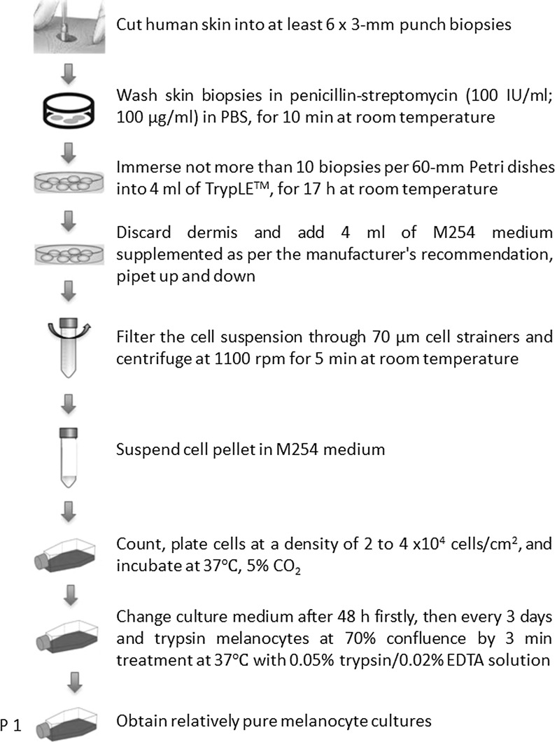
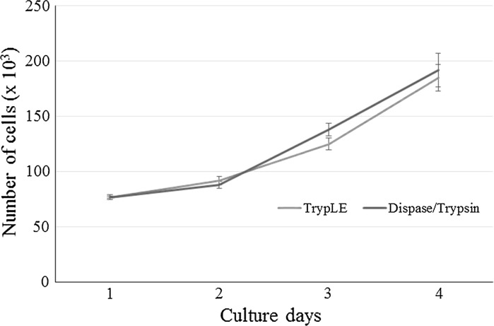
Primary melanocytes in culture are useful models for studying epidermal pigmentation and efficacy of melanogenic compounds, or developing advanced therapy medicinal products. Cell extraction is an inevitable and critical step in the establishment of cell cultures. Many enzymatic methods for extracting and growing cells derived from human skin, such as melanocytes, are described in literature. They are usually based on two enzymatic steps, Trypsin in combination with Dispase, in order to separate dermis from epidermis and subsequently to provide a suspension of epidermal cells. The objective of this work was to develop and validate an extraction method of human skin melanocytes being simple, effective and applicable to smaller skin samples, and avoiding animal reagents. TrypLE™ product was tested on very limited size of human skin, equivalent of multiple 3-mm punch biopsies, and was compared to Trypsin/Dispase enzymes. Functionality of extracted cells was evaluated by analysis of viability, morphology and melanin production. In comparison with Trypsin/Dispase incubation method, the main advantages of TrypLE™ incubation method were the easier of separation between dermis and epidermis and the higher population of melanocytes after extraction. Both protocols preserved morphological and biological characteristics of melanocytes. The minimum size of skin sample that allowed the extraction of functional cells was 6 × 3-mm punch biopsies (e.g., 42 mm2) whatever the method used. In conclusion, this new procedure based on TrypLE™ incubation would be suitable for establishment of optimal primary melanocytes cultures for clinical applications and research.

Keywords: Cell culture; Extraction; Melanocytes; Pigmentation; Skin.


Conflict of interest statement

The authors declare that they have no conflict of interest.

Figures

Fig. 1
Fig. 1
Schematic diagram for the isolation of melanocytes from small amount of human skin, via the TrypLE™ method. The initial seeding density for initiating the primary culture is 2–4 × 104 cells per 25 cm2 tissue culture flask. Generally, one such flask is set up from an initial yield of 4–10 × 105 cells isolated from about 42–70 mm2 piece of skin
Fig. 2
Fig. 2
Epidermal-dermal separation using TrypLE™ enzyme 1X (a) and Dispase II (b). After TrypLE™ incubation, epidermis (arrow) was completely separated from dermis, whereas after Dispase treatment, it was not perfectly detached
Fig. 3
Fig. 3
Skin epithelial cells isolated from multiple 3-mm punch biopsies. Comparison of number of cells using TrypLE™ and Dispase/Trypsin methods. Cell counts were performed at the extraction day and the first passage (P1). Histograms are mean ± SE of five independent experiments with skin from different donors. Whatever the method used, there was no significant difference in cell number from 6 biopsies to 10 biopsies (P > 0.05)
Fig. 4
Fig. 4
Morphology of cells extracted from 10 punch biopsies of skin, by TrypLE™ (a, c) and Dispase/Trypsin (b, c), at 2 days (a, b) and 10 days (c, d) after cell isolation. Extracted cells attached to the culture flask withindays. After 10 days of incubation, melanocyte-like morphology (dendritic cells) was dominant with TrypLE™ method whereas melanocyte and keratinocyte-like morphologies (dendritic and polygonal cells) were observed with Dispase/Trypsin method. Bar 100 µm
Fig. 5
Fig. 5
MITF expression profiles of cells extracted from 10 punch biopsies of skin, by TrypLE™ (a, c) and Dispase/Trypsin (b, c), at 2 days (a, b) and 10 days (c, d) after cell isolation. The percentage of cells expressing MITF (a marker of melanocytes) is indicated in cytometry histograms. With TrypLE™ method, 94–98% of cells were positive whereas with Dispase/Trypsin method, 53–57% of cells were positive
Fig. 6
Fig. 6
Number of viable melanocytes extracted by TrypLE and Dispase/Trypsin methods. Melanocytes at passage 2 were deposited in 12-well plates at a density of 6 × 104 cells/well. Cell numbers were measured after 1, 2, 3 and 4 days of culture. Data are expressed as the mean ± SE of 5 independent experiments carried out in duplicate. No difference was found (P > 0.05)
Fig. 7
Fig. 7
Intracellular melanin content normalized to the amount of protein. Melanocytes at passage 2 were deposited on 25 cm2 flask at a density of 4 × 105 cells/flask. Melanin and protein contents were measured after 5 days of culture. Data are expressed as the mean ± SE of 5 independent experiments carried out in duplicate. No difference was found between TrypLE and Dispase/Trypsin methods (P > 0.05)
Fig. 8
Fig. 8
Immunostaining of cells extracted from 10 punch biopsies of skin, by TrypLE™ (a) and Dispase/Trypsin (b), and cultured at passage 2. MITF (in red) was used as a characteristic melanocyte marker while F actin (green) as a cell shape marker. Cells extracted by both methods positively expressed MITF and F actin. Bar 50 µm. (Color figure online)

Similar articles

Cited by

References

    1. Duval C, Schmidt R, Regnier M, Facy V, Asselineau D, Bernerd F. The use of reconstructed human skin to evaluate UV-induced modifications and sunscreen efficacy. Exp Dermatol. 2003;12:64–70. doi: 10.1034/j.1600-0625.12.s2.10.x. - DOI - PubMed
    1. Eisinger M, Marko O. Selective proliferation of normal human melanocytes in vitro in the presence of phorbol ester and cholera toxin. Proc Natl Acad Sci USA. 1982;79:2018–2022. doi: 10.1073/pnas.79.6.2018. - DOI - PMC - PubMed
    1. Gilchrest BA, Vrabel MA, Flynn E, Szabo G. Selective cultivation of human melanocytes from newborn and adult epidermis. J Invest Dermatol. 1984;83:370–376. doi: 10.1111/1523-1747.ep12264638. - DOI - PubMed
    1. Godwin LS, Castle JT, Kohli JS, Goff PS, Cairney CJ, Keith WN, Sviderskaya EV, Bennett DC. Isolation, culture, and transfection of melanocytes. Curr Protoc Cell Biol. 2014;63:1.8.1–1.8.20. doi: 10.1002/0471143030.cb0108s63. - DOI - PubMed
    1. Guerra L, Primavera G, Raskovic D, Pellegrini G, Golisano O, Bondanza S, Paterna P, Sonego G, Gobello T, Atzori F, Piazza P, Luci A, De Luca M. Erbium: YAG laser and cultured epidermis in the surgical therapy of stable vitiligo. Arch Dermatol. 2003;139:1303–1310. doi: 10.1001/archderm.139.10.1303. - DOI - PubMed

LinkOut - more resources