. 2018:1753:217-233.
doi: 10.1007/978-1-4939-7720-8_15.

Functional and Morphological Analysis of OFF Bipolar Cells

Affiliations

Functional and Morphological Analysis of OFF Bipolar Cells

Chase B Hellmer et al. Methods Mol Biol. 2018.

Abstract

Retinal first-order neurons, photoreceptors, receive visual inputs and convert them to neural signals. The second-order neurons, bipolar cells then sort out the visual signals and encode them through multiple neural streams. Approximately 15 morphological types of bipolar cells have been identified, which are thought to encode different aspects of visual signals such as motion and color (Ichinose et al. J Neurosci 34(26):8761-8771, 2014; Euler et al. Nat Rev Neurosci 15(8):507-519, 2014). To investigate functional aspects of OFF bipolar cells, single cell recordings are preferred; however, bipolar cells in the mouse retina are small and hard to distinguish from other types of cells. Here, we describe our methodology and tips for immunohistochemistry and patch clamp recordings for analyzing light-evoked responses in each type of OFF bipolar cell.

Keywords: Immunohistochemistry; Light responses; Neurobiotin; OFF bipolar cells; Patch clamp; Retinal slice preparation.


Figures
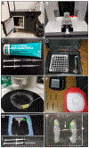
Figure 1
Figure 1
Preparation for the eye dissection. (A) A ventilated cart to place the animal cage inside for dark adaptation. (B) A handmade chopper with a razor blade (red arrow) inserted. A micrometer dial on the bottom moves the slicing chamber laterally during the dissection. (C) Grease pens are assembled from a 1 ml syringe and a pipetter tip (~200 µl). Vacuum grease is inserted into the back of the syringe. (D) A dark box for holding retinal preparations. The bottom is filled with water and oxygen is bubbled continuously. When the box is closed, the inside is completely dark and a moist, oxygenated environment is continually provided. Preparations sit on the shelf. (E) A dissecting chamber filled with HEPES solution. PE tubing from an oxygen source continuously delivers oxygen to the retinal tissue. (F) All tools and pipettes are arranged next to the dissecting microscope, with HEPES solution on ice nearby. (G) A slicing chamber to hold retina tissue (white arrow) attached to the Millipore filter paper (green arrow). (H) A plastic coverslip with parallel grease rails (yellow asterisks) and an attached retinal slice (white arrow) with filter paper (green arrow) set into the grease. (I) A micro-pipette filler and a 1 ml pipette for reference.
Figure 2
Figure 2
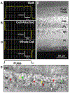
Patch clamping of OFF bipolar cells. (A) An example of the current in response to a pulse of voltage injection (1-10 mV) through the pipette when it is in the bath solution. An electrical resistance of 7-11 MΩ should be seen here. (B) An example of the current response after the giga-seal is made between the cell membrane and the pipette. The transient current at the beginning and the end of the voltage-pulse is sharp and skinny. (C) An example of the current response after the membrane has been ruptured and the whole cell configuration has been achieved. The transient is large and wider compared to the current with the “Cell Attached” configuration (B). (D) A DIC image of the entire mouse retinal slice. The photoreceptors, nuclear layers and plexiform layers are indicated to the right. (E) An enlarged DIC image of the INL. OFF bipolar cells are marked with red arrows, amacrine cells are marked with white arrows, and rod bipolar cells are marked with green arrows. Figure 2E is reproduced from (Hellmer and Ichinose, 2015) [23] with permission from the Journal of Visualized Experiments (JoVE). Abbreviations: ONL – outer nuclear layer; OPL – outer plexiform layer; INL – inner nuclear layer; IPL – inner plexiform layer; GCL – ganglion cell layer
Figure 3
Figure 3
Immunostaining of OFF bipolar cells (BC). (A) Axon terminals of type-1 BC ramify in the outer portion of the IPL, above the OFF ChAT band, and are not labeled with Syt2B. (B) Axon terminals of type-2 BC ramify to a similar portion of the IPL, above the OFF ChAT band, and is positive for Syt2B. (C) Axon terminals of type 3 BC ramify below the OFF ChAT band. (D) Axon terminals of type 4 BC ramify diffusely through the sublamina a. (E) A sulforhodamine B-filled cell seen in real time during patch clamping. (F) A type 3a BC is positive for HCN4. (G) A type 3b BC is positive for PKARIIβ. Figure 3 is reproduced from (Ichinose and Hellmer, 2016) [22] with permission from John Wiley & Sons, Inc. [Copyright © 1999-2017 John Wiley & Sons, Inc.].

References

    1. Ichinose T, Fyk-Kolodziej B, Cohn J (2014) Roles of ON cone bipolar cell subtypes in temporal coding in the mouse retina. J Neurosci 34 (26):8761–8771. doi:10.1523/JNEUROSCI.3965-13.2014 - DOI - PMC - PubMed
    1. Euler T, Haverkamp S, Schubert T, Baden T (2014) Retinal bipolar cells: elementary building blocks of vision. Nat Rev Neurosci 15 (8):507–519 - PubMed
    1. Wu SM, Gao F, Maple BR (2000) Functional architecture of synapses in the inner retina: segregation of visual signals by stratification of bipolar cell axon terminals. J Neurosci 20 (12):4462–4470 - PMC - PubMed
    1. Ghosh KK, Bujan S, Haverkamp S, Feigenspan A, Wässle H (2004) Types of bipolar cells in the mouse retina. J Comp Neurol 469 (1):70–82 - PubMed
    1. Wässle H, Puller C, Muller F, Haverkamp S (2009) Cone contacts, mosaics, and territories of bipolar cells in the mouse retina. J Neurosci 29 (1):106–117 - PMC - PubMed

MeSH terms

LinkOut - more resources