

Spontaneous intracranial vertebral artery dissection with acute ischemic stroke: High-resolution magnetic resonance imaging findings
- PMID: 29565222
- PMCID: PMC5958508
- DOI: 10.1177/1971400918764129
Spontaneous intracranial vertebral artery dissection with acute ischemic stroke: High-resolution magnetic resonance imaging findings
Abstract
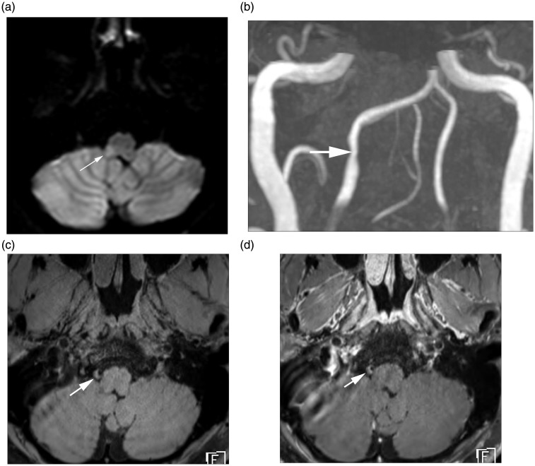
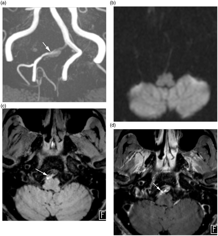
Background Acute ischemic stroke (AIS) more frequently develops in patients with intracranial vertebral artery dissection (VAD) than extracranial VAD, and is associated with possible poor clinical outcomes. The aim of this study is to compare high-resolution magnetic resonance imaging (HR-MRI) findings and clinical features of VAD with and without AIS. Methods Twenty-nine lesions from 27 patients (15 male and 12 female patients; age range = 28-73 years) who underwent diffusion MRI and 3T HR-MRI within seven days were included. We classified VAD according to the presence of AIS lesions on diffusion MRI. Clinical features and HR-MRI findings (angiographic patterns, presence of double lumen sign, dissecting flap, posterior inferior cerebellar artery involvement, remodeling index, length of affected vessels, T1-signal intensity, area of intramural hematoma, and grades and patterns of vessel wall enhancement) were evaluated. Results Thirteen VADs with AIS and 16 without AIS were included. There were no significant differences in the clinical parameters (sex, age, risk factors, symptoms). More VADs with AIS presented as a steno-occlusive pattern than VADs without AIS. More VADs without AIS presented with aneurysmal dilation, larger mean remodeling index and longer mean length than VADs with AIS. Presence of intramural hematoma, T1-iso-signal intensity of intramural hematoma and contrast enhancement were significantly more common in VADs with AIS than without AIS. Conclusions Our study showed some differences in HR-MRI comparing intracranial VAD patients with and without AIS. Differing findings may facilitate a better understanding of intracranial VAD and risk assessment of AIS in these patients.
Keywords: Acute ischemic stroke (AIS); high-resolution magnetic resonance imaging (HR-MRI); intramural hematoma; vertebral artery dissection (VAD).
Figures
Similar articles
-
Intramural hematoma detection by susceptibility-weighted imaging in intracranial vertebral artery dissection.Cerebrovasc Dis. 2013;36(4):292-8. doi: 10.1159/000354811. Epub 2013 Oct 16. Cerebrovasc Dis. 2013. PMID: 24135546
-
Utility of T1- and T2-Weighted High-Resolution Vessel Wall Imaging for the Diagnosis and Follow Up of Isolated Posterior Inferior Cerebellar Artery Dissection with Ischemic Stroke: Report of 4 Cases and Review of the Literature.J Stroke Cerebrovasc Dis. 2017 Nov;26(11):2645-2651. doi: 10.1016/j.jstrokecerebrovasdis.2017.06.038. Epub 2017 Aug 30. J Stroke Cerebrovasc Dis. 2017. PMID: 28864037 Review.
-
Monitoring Intramural Hematoma on Vessel Wall Imaging to Evaluate the Healing of Intracranial Vertebral Artery Dissection.J Stroke Cerebrovasc Dis. 2021 Sep;30(9):105992. doi: 10.1016/j.jstrokecerebrovasdis.2021.105992. Epub 2021 Jul 19. J Stroke Cerebrovasc Dis. 2021. PMID: 34293642
-
Utility of cone-beam computed tomography angiography for the assessment of vertebral artery dissection.J Clin Neurosci. 2018 Feb;48:76-80. doi: 10.1016/j.jocn.2017.11.010. Epub 2017 Nov 26. J Clin Neurosci. 2018. PMID: 29257748
-
Magnetic resonance imaging, magnetic resonance and catheter angiography for diagnosis of cervical artery dissection.Front Neurol Neurosci. 2005;20:102-118. doi: 10.1159/000088155. Front Neurol Neurosci. 2005. PMID: 17290116 Review.
Cited by
-
A Case of Intracranial Vertebral Artery Dissection Undetected by CT, MRI, and MRA at the Onset of Headache That Caused Subarachnoid Hemorrhage Seven Days Later.J Neuroendovasc Ther. 2022;16(5):265-269. doi: 10.5797/jnet.cr.2021-0033. Epub 2021 Sep 11. J Neuroendovasc Ther. 2022. PMID: 37502233 Free PMC article.
-
Image Findings of Acute to Subacute Craniocervical Arterial Dissection on Magnetic Resonance Vessel Wall Imaging: A Systematic Review and Proportion Meta-Analysis.Front Neurol. 2021 Apr 7;12:586735. doi: 10.3389/fneur.2021.586735. eCollection 2021. Front Neurol. 2021. PMID: 33897578 Free PMC article.
-
Quantitative evaluation for intravascular structures of vertebral artery dissection with a novel zoomed high-resolution black-blood MR imaging.Neuroradiol J. 2023 Oct;36(5):563-571. doi: 10.1177/19714009231163557. Epub 2023 Mar 14. Neuroradiol J. 2023. PMID: 36916331 Free PMC article.
-
Top of basilar syndrome due to vertebral artery dissection: How high-resolution MRI and CD31 analysis of thrombus could help.Int J Surg Case Rep. 2023 Nov;112:108948. doi: 10.1016/j.ijscr.2023.108948. Epub 2023 Oct 10. Int J Surg Case Rep. 2023. PMID: 37832359 Free PMC article.
-
MR Intracranial Vessel Wall Imaging: A Systematic Review.J Neuroimaging. 2020 Jul;30(4):428-442. doi: 10.1111/jon.12719. Epub 2020 May 11. J Neuroimaging. 2020. PMID: 32391979 Free PMC article.
References
-
- Bachmann R, Nassenstein I, Kooijman H, et al. Spontaneous acute dissection of the internal carotid artery: high-resolution magnetic resonance imaging at 3.0 Tesla with a dedicated surface coil. Invest Radiol 2006; 41: 105–111. - PubMed
-
- Bogousslavsky J, Regli F. Ischemic stroke in adults younger than 30 years of age: cause and prognosis. Arch Neurol 1987; 44: 479–482. - PubMed
-
- Takano K, Yamashita S, Takemoto K, et al. MRI of intracranial vertebral artery dissection: evaluation of intramural haematoma using a black blood, variable-flip-angle 3D turbo spin-echo sequence. Neuroradiology 2013; 55: 845–851. - PubMed
-
- Kristensen B, Malm J, Carlberg B, et al. Epidemiology and etiology of ischemic stroke in young adults aged 18 to 44 years in Northern Sweden. Stroke 1997; 28: 1702–1709. - PubMed
-
- Kim B, Kim S, Kim D, et al. Outcomes and prognostic factors of intracranial unruptured vertebrobasilar artery dissection. Neurology 2011; 76: 1735–1741. - PubMed
MeSH terms
LinkOut - more resources
Full Text Sources
Other Literature Sources
Medical
